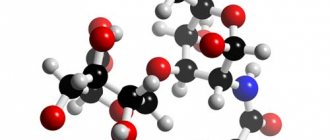
Phosphatidylcholines (Pcholines), whose formula is shown in the picture below, are a class of phospholipids that include choline as a head group.
They are the main component of biological membranes. Can be easily obtained from various readily available sources such as egg yolk or soybeans, from which they are extracted mechanically or chemically using hexane. They are also part of the lecithin group of yellow-brown fatty substances found in animal and plant tissues. Dipalmitoylphosphatidylcholine (eg, lecithin) is a major component of pulmonary surfactant and is often used in the L/S ratio to calculate fetal lung maturity. Although these substances are found in all plant and animal cells, they are absent from the membranes of most bacteria, including Escherichia coli. They are produced commercially in purified form.
You might be interested in: What is a gallery?
Etymology
The name "lecithin" was originally determined from the Greek "lekyte" (λεκιθος, i.e. egg yolk) by Théodore Nicolas Gobley, a mid-19th century French chemist and pharmacist who applied it to egg yolk phosphatidylcholine. This is what he identified in 1847.
You may be interested in: A spiteful critic is a person with a deliberately bad attitude
Gobley eventually fully described lecithin from a chemical structural point of view in 1874. In some contexts, the terms phosphatidylcholine/lecithin are used interchangeably. However, lecithin extracts consist of a mixture of F. and other compounds. It is also used in conjunction with sodium taurocholate to simulate biorelevant fed and fasted environments in dissolution studies of highly lipophilic drugs.
Localization
Phosphatidylcholine formula is a major component of cell membranes and pulmonary surfactant and is more commonly found in the exoplasmic or outer envelope of the cell membrane.
F. also plays a role in membrane-mediated cell signaling and PCTP activation of other enzymes.
You might be interested in: Social studies about society and nature. Consequences of scientific and technological progress
The phospholipid phosphatidylcholine consists of a group of choline and glycerophosphoric acid with various fatty acids. It is usually a saturated fatty acid (this may be palmitic or hexadecanoic acid, H3C-(CH2)14-COOH; margaric acid, identified by Gobley in egg yolk, or heptadecanoic acid H3C-(CH2)15-COOH, which also belongs to this class) or unsaturated fatty acid (oleic, or 9Z-octadecenoic, as in Gobli's original egg yolk lecithin).
Journal "Child's Health" 6(9) 2007
Recently, many research centers have carried out fundamental and exploratory work aimed at a comprehensive study of extensive natural biologically active compounds, united under the general name “lipids”. The idea of lipids as a passive depot for supplying cells with energy is a thing of the past. Modern ideas, based on the results of in-depth structural and functional studies, assign lipids and their supramolecular cellular formations—biological membranes—the most important role in the functioning of the basic biochemical mechanisms that determine and regulate the physiological state of the cell, its reactions and interactions with both neighboring cells and environmental factors [12, 16].
Certain types of natural lipid substances, their biologically active molecular fragments and modified synthetic analogs attract great attention from researchers as a promising source for the design of new generation drugs and diagnostics [7, 32, 34].
Among the many natural lipids, the most significant are phospho- and glycophospholipids. Phospholipids are present in the largest quantities in membranes, which serve as their structural components and are never stored in the body in large quantities. Phospholipids, although in small quantities, are found in the cells of organs and tissues of all types of living organisms, and increased concentrations of these natural substances are observed in the brain and spinal cord, heart muscle, liver, and lungs.
Data from X-ray diffraction analysis and others show that phospholipid molecules have the shape of a laterally flattened cylinder, and are divided along the length into two unequal parts: a small “head” consisting of polar groups and a long “tail” formed by hydrocarbon chains of fatty acids, included in the phospholipid. In phospholipids, the “head” consists of sequentially interconnected residues of a nitrogenous base (choline, colamine or serine), a phosphate group and glycerol. Fatty acid residues forming hydrophobic “tails” are connected to glycerol. The saturated acid is usually palmitic, and the unsaturated acid is oleic, linoleic, linolenic, etc. The same compound, but without a nitrogenous base, is called phosphatidic acid; in its free form it is found in very small quantities, playing the role of an intermediate product in the synthesis of phospholipids. Membrane asymmetry of phospholipids is a universal phenomenon characteristic of almost all cells [17]. It has now been established that the maintenance of asymmetry is an active process realized through the activity of ATP and the sulfhydryl-dependent lipid pump (aminophospholipid translocase), which moves aminophospholipids towards the inner membrane [25]. Activation leads to increased movement of phospholipids between layers, which is accompanied by loss of asymmetry [36]. In particular, platelets, when stimulated, lose their normal membrane asymmetry, which leads to an increase in the expansion of anionic PS. This process plays an important physiological role in the development of the local blood coagulation reaction [25, 36]. In addition, the surface exposure of PS (at least on erythrocytes) is a signal for the rapid removal of these cells from the bloodstream.
Negatively charged phospholipids create a surface on which the assembly of enzyme complexes of the two main reactions of the coagulation cascade occurs. In one of them (the tenase complex), factor X is activated by a complex of factors IXa and VIIIa, and in the other (prothrombinase complex), a prothrombinase reaction occurs, the conversion of prothrombin to thrombin by an enzyme complex consisting of factors Xa and Va. The interaction of factors IXa, Xa and prothrombin with the lipid surface occurs through the formation of a calcium-dependent bridge between the gamma-carboxyglutamic acid residues of these proteins and the negatively charged polar groups of phospholipids [38]. Binding to the lipid surface leads to an increase in local concentration and more efficient location of coagulation factors, which contributes to the maximum rate of reaction. Any substances that interfere with the assembly of these complexes on the phospholipid surface, including antibodies to phospholipids, have the potential to increase the level of thrombin formation and impair blood coagulation [37].
Phospholipids are usually considered to be derivatives of phosphatidic acid. This is where the name of a number of important phospholipids comes from, in particular their main representative, phosphatidylcholine. Cholesterol differs in structure from the first three classes of lipids. It is an extended system of four hydrocarbon rings and a hydrocarbon side chain and is hydrophobic except for one hydroxyl group, which forms a polar "head". Cholesterol is part of the outer cell membrane and forms a rigid structure, impairing such properties of the membrane as fluidity. Each type of membrane has a strictly defined content of various classes of lipids, which largely determines its properties [21, 25].
With an increase in the content of phospholipids in the membrane, its lability increases, which facilitates various types of diffusion. As a rule, internal membranes are more labile, i.e. they are more fluid and more permeable [16, 17]. Thus, phospholipids provide the basic properties of cell membranes; they are endogenous compounds, the synthesis of which is disrupted when the cell is damaged. This contradiction can be overcome by introducing these substances from the outside, in which case they are called essential phospholipids (EPL). EPLs are highly purified phospholipid extracts obtained from soybeans containing unsaturated fatty acids. There are two degrees of purification of EPL - with 72–76% and 92–96% phosphatidylcholine content. Of primary therapeutic importance is a compound called 1,2-dilinoleoylphosphatidylcholine (DLPC), which is an integral part of the drug and has a number of beneficial properties [4, 10].
Essential phospholipids (EPL substance) are complex substances of natural origin, which are diglycerol esters of choline phosphoric acid (phosphatidylcholine) and unsaturated fatty acids (linoleic, linolenic, oleic). Preparations of essential phospholipids have membranotropic properties; metabolic and hepatoprotective effects; regulate lipid and carbohydrate metabolism. The most studied use of EPL is in the treatment of liver diseases [4, 10, 11].
Essential phospholipids are the main elements in the structure of the cell membrane and cellular organelles (mitochondria). In the mechanism of hepatoprotective action, an important role is played by the replacement effect [33]. When liver metabolism is disrupted, essential phospholipids provide the supply of high-energy phospholipids, ready for absorption, which are ideally combined with endogenous phospholipids in chemical structure. They mainly penetrate liver cells, penetrating their membranes [32]. Treatment with essential phospholipids leads to a significant improvement in biochemical parameters such as serum ALT, AST, as well as a decrease in necrosis and inflammation. Positive dynamics of indicators (reduction of intralobular necrosis, portal inflammation, general improvement in well-being) compared to the initial level was observed in patients taking essential phospholipid preparations for one year [27, 28]. The spectrum of activity of essential phospholipids in chronic degenerative liver diseases (CDLD) can be presented as follows: restoration and preservation of the integrity of hepatocyte membranes; activation of membrane phospholipid-dependent enzymes; improvement of lipid metabolism during the synthesis of lipoproteins in the liver; activation of RNA synthesis, normalization of protein metabolism; increased glycogen content in the liver; increasing detoxification excretory potential (detoxification function of the liver); converting neutral fats and cholesterol into easily metabolized forms; reducing the level of energy expenditure of the liver; reduction and disappearance of fatty infiltration of hepatocytes; antifibrotic effects (reducing the risk of connective tissue development: fibrosis and cirrhosis of the liver); stabilization of the physicochemical properties of bile [11, 32].
The good tolerability of essential phospholipids allows us to consider them as a fairly powerful means for increasing the body's resistance, which allows us to significantly expand the indications for their use. Essential phospholipids are well absorbed in the small intestine, including in the form of biologically active products of their hydrolysis by digestive enzymes, which are transported through the intestinal wall into the lymphatic channel. Partial resynthesis of phosphatidylcholine from hydrolysis products occurs in the liver.
Essential phospholipids are widely used as part of complex therapy for the following conditions: hepatitis (acute and chronic), toxic hepatitis, drug and alcohol liver damage (alcoholic hepatitis), poisoning; fatty liver - fatty hepatosis of various origins (diabetes mellitus, chronic infections); liver dysfunction in somatic diseases; cirrhosis of the liver; liver cell necrosis, liver failure, hepatic coma and precoma (solution for injection); pre- and postoperative treatment, especially during operations in the hepatobiliary zone, such as cholecystectomy (solution for injection); hyperlipoproteinemia, hypercholesterolemia, hypertriglyceridemia; diseases of the cardiovascular system: coronary heart disease, angina pectoris, condition after myocardial infarction and stroke, cerebral and peripheral circulatory disorders, hypertension, vascular atherosclerosis, diabetic angiopathy, prevention of thromboembolism before surgery, prevention and treatment of fat embolism (solution for injection ); diseases of the digestive system (chronic pancreatitis, peptic ulcer of the stomach and duodenum, etc.); toxicosis of pregnancy (edema, proteinuria and hypertensive disorders); radiation sickness (radiation syndrome); psoriasis, atopic dermatitis, diffuse neurodermatitis, eczema; prevention of premature aging [1, 2, 15, 18, 20, 23, 26–28, 35].
Liposomes are artificially created lipid vesicles (vesicles) consisting of one or more phospholipid bilayers separated by an aqueous phase. The diameter of liposomes can range from 25 to 10,000 nm. In the mid-60s, the English scientist Alec Bangham, elucidating the role of phospholipids in blood clotting, studied the structure of dispersions formed when phospholipids swell in excess water. In electron micrographs, he saw layered particles similar to the membrane structures of a cell. The next study showed that the elements present in the solution at the time of phospholipid swelling are included inside these particles and are retained there for a long time, exchanging with the outer solution at a very low rate. This was the first time it was established that phospholipids, which are the main components of cell membranes, are capable of spontaneously forming closed membranes in water. These shells capture part of the surrounding aqueous solution, and the phospholipid membrane that forms them has the properties of a semi-permeable barrier that easily allows water to pass through, but prevents the penetration of substances dissolved in it. The properties of liposomes and their behavior are determined primarily by the presence of a closed membrane shell (Fig. 1). Despite its molecular thickness (about 4 nm), the lipid bilayer is characterized by exceptional mechanical strength and flexibility. Thanks to this, liposomes maintain their integrity under various damaging influences, and their membrane has the ability to self-heal structural defects that arise in it. At the same time, the flexibility of the bilayer and its fluidity give liposomes high plasticity [8, 9, 14, 19]. Typically, liposomes are prepared by shaking or sonicating aqueous suspensions of phospholipids. Liposomes can be formed from individual phospholipids, both natural and synthetic, as well as from a mixture of phospholipids [7, 12].
Many drugs have a low therapeutic index. This means that the concentration at which they have a therapeutic effect differs little from the concentration at which the drug becomes toxic. In other cases, a drug may quickly lose activity when introduced into the body. The inclusion of such drugs in liposomes can significantly increase their therapeutic effectiveness, since, on the one hand, the drug located in the liposome is protected by its membrane from the action of unfavorable factors, and on the other hand, the same membrane does not allow the toxic drug to exceed the permissible concentration in biological fluids of the body . In this case, the liposome acts as a storage facility from which the drug is released gradually, in the required doses and over the required period of time [6, 13, 14].
Improving the quality of medical care for children at the present stage of scientific development is only possible with the introduction of modern advanced technologies for the production of new, effective dosage forms of drugs.
The use of liposome-drug complexes has a number of advantages over the use of drugs alone: liposomes allow the delivery of substances into cells that, in the absence of liposomes, do not penetrate into them; attachment of appropriate antibodies (vectors) to liposomes can ensure delivery of substances to target cells; a drug encapsulated in liposomes provides a greater therapeutic effect (the duration of action increases, and the dose can be significantly reduced); liposomes can be effectively used as adjuvants, i.e. substances that stimulate immunological reactions [14, 24]. It is assumed that there are at least 2 ways for liposomes to enter the cell (Fig. 2).
The first is that, due to endocytosis, the liposome is taken up by the cell and a vacuole is formed, which fuses with lysosomes. Phospholipases of lysosomes hydrolyze phospholipids of the lysosome membrane, ensuring the release of the drug into the cytoplasm of the cell. If the liposome consists of several lipid membranes, then their gradual hydrolysis ensures a slow release of the drug into the cell. The second way is when liposomes fuse with the cell membrane, whereby the lipid component of the liposome is incorporated into the cell membrane, and the water-soluble drug penetrates the cytoplasm. Thus, in both cases, the substance encapsulated in the liposome enters the cells despite the membrane barrier. Experiments on animals have shown that with intravenous, intramuscular and intraperitoneal administration, liposomes quite quickly leave the bloodstream, as they are captured by the cells of the macrophage system, primarily by the cells of the liver and spleen [21].
The liposomal form of natural phosphatidylcholine is the drug lipin, which is a lyophilized form of liposomes from egg phosphatidylcholine (Fig. 3).
An experimental study of the pharmacokinetics of liposomes from phosphatidylcholine when administered intratracheally into the body, carried out on intact rats and on a model of acute pneumonia using radioactive markers, showed that the drug is distributed fairly quickly and evenly in the lungs. This type of administration ensures a long stay of liposomes in the lungs - up to 6 hours, and in animals with a model of acute pneumonia, liposomes remained in the lungs in greater quantities than in animals in the control group. At the same time, the authors found that a significant part of the drug is retained in the liver and kidneys, and the content of liposomes in these organs in control rats is significantly higher than in animals with acute pneumonia. Experimental work on the use of lipin on experimental models of induced liver dystrophy, which is accompanied by an increase in the amount of primary and secondary products of lipid peroxidation in homogenates, a decrease in the activity of antioxidant enzymes, showed that the use of lipin in combination with vitamin E, selenium and cordiamine has a positive effect on the condition liver. The indicators of lipid peroxidation in animals that were administered lipin in combination with vitamin E and lipin in combination with selenium were almost completely normalized. Under the influence of the drugs, the secretory function of the liver was completely normalized. It has been established that the use of lipin normalizes the content of micro- and macroelements of lung surfactant in experimental animals. At the same time, it has been shown that lipin is more effective when used early after the damaging effect of sulfur dioxide administered by inhalation - after 5 minutes, and to a lesser extent - after 24 hours. The use of lipin after 7 days was ineffective. The possibility of correction of central hemodynamic disturbances during hemorrhagic shock using lipin was demonstrated, which allowed the authors to conclude that phosphatidylcholine liposomes have an antihypoxic and antioxidant effect. The introduction of liposomes into the bloodstream helps restore the functional activity of cell membranes damaged due to hypoxia, and primarily the endothelium of blood vessels [1, 5, 22, 26, 28].
There is information about the effectiveness of lipin use in children with bronchial asthma. It was shown that lipin administered using ultrasonic inhalation contributed to the normalization of most parameters of respiration and circulation, and optimization of the relationship between ventilatory and hemodynamic parameters in the lungs [22, 24, 27].
When using lipin in patients with purulent-destructive lung diseases complicated by respiratory failure syndrome, it was found that already 1 hour after inhalation administration of the drug there was a statistically significant increase in the vital capacity of the lungs, the dynamics of the blood gas composition is characterized by a tendency to increase the partial pressure of oxygen and oxygen saturation arterial blood [23, 35].
The antioxidant and antihypoxic activity of lipin was studied when used in patients with broncho-obstructive diseases complicated by respiratory failure. It was found that already after the first inhalation there is a tendency towards a decrease in the activity of lipid peroxidation with a simultaneous increase in the activity of antioxidant enzymes (superoxide dismutase and catalase). In addition, a decrease in the activity of aminotransferases and a decrease in the amount of pyruvic acid were found, which indicates the normalization of the glycolysis process [23]. The data presented indicate that lipin has an antihypoxic effect, facilitating an increase in the rate of diffusion of oxygen from the lungs into the blood and from the blood into the tissues, and normalizes the processes of tissue respiration. The drug restores the functional activity of endothelial cells, the synthesis and release of endothelial relaxation factor (NO), improves microcirculation and rheological properties of blood. It inhibits the processes of lipid peroxidation in the blood and tissues, maintains the activity of the body's antioxidant properties, has a membrane protective effect, and increases nonspecific immunity. When administered by inhalation, lipin helps preserve pulmonary surfactant, which, in turn, improves pulmonary and alveolar ventilation and increases the rate of oxygen transport through biological membranes. The drug does not disrupt the functional activity of organs and systems of the body and is non-toxic.
Due to the fact that in 2007 Ukraine switched to the international statistical system in accordance with WHO recommendations, pediatric neonatologists were tasked with caring for newborns with low, very low and extremely low birth weight. The most common pathology that determines high morbidity and mortality in this population is respiratory distress syndrome (RDS). The development of neonatal resuscitation leads to an increase in the number of premature newborns who survive after prolonged ventilation support, who subsequently constitute a risk group for the formation and development of chronic lung diseases [45, 66, 68–70]. The basis for the development of RDS in newborns is the structural and functional immaturity of the lungs and the surfactant system [46]. In 1959, Avery and Mead were the first to demonstrate surfactant deficiency in children who died from RDS. They noted that surface tension forces in the lungs of children who died from hyaline membrane disease were much higher than those who died from other causes, and established an inverse relationship between these forces and the gestational age of the child. Surfactant is a monomolecular layer at the interface between the alveolar epithelium and the air and is a lipoprotein. 90% of the surfactant consists of lipids, mainly phospholipids (PL): dipalmitoylphosphatidylcholine (DPPC) - 45%, phosphatidylcholine (PC) - 25%, phosphatidylglycerol (PG) - 5%, remaining phospholipids - 5%, other lipids (cholesterol, triglycerides, unsaturated fatty acids, sphingomyelin) - 10%. The remaining 10% falls on the protein fraction, which is represented by apoproteins. Four surfactant-associated proteins (apoproteins) have been identified: SP-A, SP-B, SP-C, SP-D, synthesized by epithelial cells of the respiratory tract and Clara cells. Apoprotein SP-A plays an important role in the metabolism of surfactant, regulates its secretion, protects against bacterial endotoxins, herpes simplex viruses, influenza, increases the phagocytic activity of macrophages and the production of free oxygen radicals [50, 51, 61, 64, 65]. Apoproteins SP-B and SP-C serve for adsorption and distribution of phospholipids in the alveolar space and the formation of tubular myelin. Apoprotein SP-D takes an active part in protecting the lungs from pathogenic microorganisms. These four surfactant proteins play an important role in increasing resistance to albumin and fibrinogen as they leak into the alveoli [40, 54]. Hereditary or congenital deficiency of surfactant protein B is inherited in an autosomal recessive manner and leads to death. It is manifested by the development of the clinical manifestations of RDS in full-term newborns with a long-term need for mechanical ventilation; replacement therapy with exogenous surfactants has only a temporary effect [59, 60].
Surfactant is synthesized by type 2 alveolocytes, which develop from the cuboidal epithelium of the distal respiratory tract from the 20–24th week of gestation, but the most active synthesis occurs starting from the 34th week. The secretion of surfactant components is carried out by exocytosis. There is active turnover between intracellular and extracellular surfactant, with the result that 80–90% of phospholipids can be recycled and reused within a few days. This makes it possible to effectively maintain surfactant function [57]. There are two routes for the synthesis of the main phospholipid component - phosphatidylcholine (lecithin): methylation of phosphatidylethanolamine using methyltransferase (the source of the methyl group is methionine) and synthesis from cytidine diphosphate choline, which reacts with diglyceride, in the presence of phosphocholine transferase [58]. Until the 33rd–35th week of gestation, surfactant synthesis is mainly carried out along the first pathway, and later - with the help of phosphatidylcholine transferase. The main link in the pathogenesis of RDS is the primary deficiency of the surfactant system, which develops in children born before 35–36 weeks of intrauterine development [52, 56]. The available reserves of surfactant ensure the onset of breathing and the formation of functional residual capacity of the lungs, but due to the lag of synthesis from the rate of breakdown of surfactant, its insufficiency occurs, which leads to collapse of the alveoli during exhalation, continuous gas exchange does not occur in the lungs, which leads to the occurrence of hypoxemia and hypercapnia. In addition, secondary disruption of the surfactant system is of great importance, leading to decreased synthesis or increased degradation of phosphatidylcholines [61]. Such factors are intrauterine or postnatal hypoxia, birth asphyxia, hypoventilation, acidosis, and infectious problems. Diabetes mellitus in the mother, birth by cesarean section, male gender, birth as the second of twins, and isoserological incompatibility of the blood of mother and fetus also predispose to the development of RDS [47, 48]. With RDS, an imbalance develops between the components of the phospholipid fraction - the amount of saturated phosphatidylcholine (lecithin) decreases and the content of phosphatidylserine and sphingomyelin increases [49]. Insufficient synthesis and rapid inactivation of surfactant lead to a decrease in lung compliance, which, in combination with reduced chest compliance in premature newborns, leads to the development of hypoventilation and inadequate oxygenation. Hypercapnia, hypoxia, and respiratory acidosis develop. This, in turn, leads to an increase in pulmonary vascular resistance with subsequent shunting of blood, both intrapulmonary and extrapulmonary, through the fetal communications. Increased surface tension in the alveoli causes their expiratory collapse with the development of atelectasis and hypoventilation zones, which further disrupts the ventilation-perfusion relationship in the lungs, exacerbating blood shunting [46]. A decrease in pulmonary blood flow, in turn, leads to ischemia of alveolocytes and vascular endothelium, which causes changes in the airborne barrier with transudation of plasma proteins into the interstitial space and the lumen of the alveoli. Children with RDS often develop ventricular myocardial dysfunction associated with ischemia of the subendocardial layer and papillary muscles of the ventricle, which may be due to the anatomical and physiological immaturity of the myocardium and its ischemic damage in the perinatal period [39, 40]. A modern method of treating RDS in premature newborns is replacement therapy with exogenous surfactants [39, 41–44, 63]. In recent years, the use of natural phosphatidylcholine preparations as surfactant-protective therapy has attracted special attention.
E.S. Keshichan et al. (1995) conducted a clinical study to determine the effectiveness of the drug lipin in newborns after prolonged artificial ventilation. The drug was administered by inhalation for 5 minutes 2 times a day with an interval of 5 hours. The drug was used in newborns who underwent artificial ventilation for 5 days. The dose of the drug was selected empirically, based on the maximum clinical effect. The effectiveness of the drug was determined by the normalization of blood gas composition, oxygen saturation, changes in clinical parameters in a daily analysis during the use of lipin and placebo (0.9% sodium chloride solution), and a comparison was made of the general clinical course of the disease (pneumonia) when using this type treatment and without it. The clinical effect of inhaled use of lipin was noted after 2-3 inhalations. The need for oxygen supplementation was reduced. On the 3rd day, all children showed an improvement in respiratory conductivity. The authors also noted the presence of a bronchodilator effect and an improvement in cough productivity. As a result of the use of this type of treatment, the duration of antibiotic therapy was reduced [15].
E.F. Cherniy (1995) used lipin, which was administered by inhalation at a dose of 10–100 mg/kg (depending on gestational age and clinical manifestations) for hypoxia in newborns in the early neonatal period. Research results have shown that the use of lipin helps to optimize external respiration, normalize ventilation-perfusion ratios, and reduce metabolic acidosis and hypoxemia. It was found that lipin has a positive effect on hemodynamic parameters, reducing heart rate, and normalizes blood pressure. As a result of the use of lipin, according to the same author, indicators of cellular adaptation in newborns with manifestations of hypoxia are improved, which leads to an improvement in the general condition and a decrease in the manifestations of respiratory failure [29–31].
Our own experience with the use of lipin allows us to talk about the possible surfactant-protective (both exogenous and baby surfactant) effect of the drug, since its use in premature newborns with RDS reduces the duration of neonatal stay on mechanical ventilation and reduces dependence on high levels of oxygen concentration. Also, with the endotracheal use of lipin, the likelihood of repeated administration of exogenous surfactant preparations is reduced, which, in addition to its positive clinical significance, has a certain financial significance. The use of inhaled lipin improves the drainage function of the tracheobronchial tree. The transfer of newborns to assisted ventilation is carried out earlier, the duration of mechanical ventilation is reduced, which, in turn, prevents the risk of developing chronic lung diseases in newborns [2, 3].
The literature data presented in this article allows us to hope for the real possibility of widespread use of liposomal forms of natural phosphatidylcholine for the treatment of diseases in the practice of a pediatrician.
Catalysis
Phospholipase D catalyzes the hydrolysis of the formula phosphatidylcholine to form phosphatidic acid (PA), releasing the soluble choline head group into the cytosol.
F. is a neutral lipid, but it carries an electric dipole moment of about 10 D. The vibrational dynamics of phosphatidylcholine and its waters of hydration have recently been calculated from first principles.
F. is a vital substance contained in every cell of the human body. Some researchers have used mutant mouse models with severe oxidative damage as a model of accelerated aging to investigate the possible role of phosphatidylcholine supplementation as a way to slow down processes associated with aging and improve brain function and memory capacity in dementia. However, a systematic review of human clinical trials in 2009 found that there was insufficient evidence to support the use of lecithin or F. for patients with dementia. The study found that a modest benefit cannot be ruled out until further large-scale studies are conducted.
Mechanism of action
Brain cells are surrounded by a membrane, which consists of two layers: outer and inner. The outer layer of each cell contains mainly phosphatidylcholine (PC) and sphingomyelin, while the inner layer contains mainly phosphatidylserine (PS), phosphatidylinositol and phosphatidylethanolamine.
The outer layer of each cell membrane is highly permeable. But the inner layer is much less permeable. These two layers of fatty acids are in a constant state of movement, vibrating millions of times per second. This constant vibration can be considered the "ground of life." And it is the basis of everything that happens in the brain.
The amount and type of long-chain fatty acids in the diet influences the composition of these cell membranes. Cell structure and function depend on the ideal balance of fats, including cholesterol, oleic, palmitic and stearic fatty acids. And essential fatty acids such as Omega-3. Without the correct balance, the functioning of cell membranes is disrupted.
Modern nutrition does not provide the ideal balance of fatty acids (phospholipids) to maintain the integrity of brain cells. This is why people experience memory loss, slow thinking and poor decision making.
Phosphatidylcholine maintains the integrity of brain cell membranes. And is directly involved in the synthesis of acetylcholine (ACh), which is necessary for cognition, learning and memory formation.
Most phosphatidylcholine is integrated into cell membranes, but some is broken down into choline and the phosphatidyl group. Choline enhances nerve transmission by enhancing the function of acetylcholine. The phosphatidyl group is involved in the synthesis of sphingomyelin, which, like myelin, is necessary for the central nervous system. Sphingomyelin deficiency contributes to systemic neurodegeneration.
Advantages
Research has examined the potential benefits of the phosphatidylcholine structural formula for liver repair. The results were conducted on animals and no clinical data suggest a benefit to human health. One study showed the healing effect of F. on mice with hepatitis A, B and C. Administration of F. in chronic active hepatitis led to a significant decrease in disease activity in rodents.
You might be interested: What is Zapravsky? Interpretation and synonyms
The danger of low PC levels
Low PC levels can lead to multiple health problems, especially age-related cognitive decline.
When cell membranes are damaged or communication between nerve cells is disrupted, problems with remembering things and learning occur. Low PC levels are believed to be a factor in the pathogenesis of several neurological diseases, including Alzheimer's disease.
Other conditions associated with low phosphatidylcholine levels:
- Schizophrenia and bipolar disorder.
- Ulcerative colitis.
- Chronic liver diseases (fatty liver).
Promotion
Some organizations promote the use of injectable fat, also known as injectable lipolysis, claiming that the procedure can break down fat cells and thus serve as an alternative to liposuction. While early experiments did not show any amount of lipolysis even remotely comparable to liposuction. Injections of phosphatidylcholine in a small number of patients have been reported to shrink or eliminate many types of lipomas, although some actually increase in size. There were side effects that went away without any complications. Long-term studies are considered necessary to evaluate effectiveness. Dr. Patrick Tracy has successfully used F. and deoxolate in the treatment of infraorbital fat pads.
What happens after the procedure (further care)
After administration of the drug, the breakdown products of fat cells enter the kidneys through the bloodstream, where they are subsequently filtered and excreted from the body naturally.
What effects does the patient receive:
- tightened, smooth skin;
- smoothed wrinkles, smooth relief;
- reduction of subcutaneous fat and the severity of folds.
During the recovery period, the patient is recommended to use various cosmetics with a lifting (since the fatty tissue will go away, the skin may lose its elasticity), anti-cellulite effect. This will help consolidate and prolong the results obtained.
Phases
A phase IIa/b clinical trial conducted at the Heidelberg University Hospital showed that delayed-release purified phosphatidylcholine is an anti-inflammatory and surface hydrophobicity agent. It has promising therapeutic potential in the treatment of ulcerative colitis.
In a 2011 report, microbial phosphatidylcholine catabolites were associated with increased atherosclerosis in mice through the production of choline, trimethylamine oxide, and betaine.
Although there are more pathways for phosphorus biosynthesis, one of them predominates in eukaryotes. It involves a condensation reaction between diacylglycerol (DAG) and cytidine-5′-diphosphocholine (CDP-choline or citicoline), mediated by the enzyme diacylglycerol choline phosphotransferase. Another prominent pathway in some tissues (mainly the liver) is the stepwise methylation of phosphatidylethanolamine with S-adenosylmethionine (SAM) as a methyl group donor.
In cells
Phosphatidylcholine is a key component of our cells. The supplement may improve mental, liver and intestinal health, protect nerves and improve memory. F. injections are also used to reduce fat. Find out more about its benefits, dosage and side effects.
Phosphatidylcholine levels may decrease with age. For example, in the brain there is a decrease of 10% between 40 and 100 years.
Since choline is required for the production of phosphatidylcholine, low choline levels can limit its production. Its deficiency can reduce the level of phosphatidylcholine in the liver, which leads to liver failure. Phosphatidylcholine is also responsible for the production of very low-density lipoprotein (VLDL) [R, R].
Low levels of F are associated with memory loss and Alzheimer's disease. A study (DB-RCT) of 80 healthy young adults found that supplementation with the lipolytic phosphatidylcholine improved memory.
F. increases choline and acetylcholine levels in the brain, improves memory and protects the brain in mice with dementia.
Very low levels of phosphatidylcholine deoxycholate can cause liver damage and even death in mice. Animal studies have shown that F. can promote liver regeneration.
Low levels of choline and phosphatidylcholine-phosphatidylserine can cause non-alcoholic fatty liver disease (NAFLD) in humans.
Side effects (10 troubles)
If the patient is being treated for various cognitive impairments with acetylcholinesterase inhibitors, then administration of phosphatidylcholine is prohibited.
This combination leads to an excessive increase in acetylcholine levels, which is fraught with cholinergic side effects:
- difficulty urinating due to bladder atony;
- constipation due to intestinal atony;
- decreased secretion by the mucous membranes and sweat glands, resulting in dry mouth, decreased sweating, and “dry” conjunctivitis;
- headache, dizziness;
- blurred vision, double vision, increased intraocular pressure;
- tachycardia;
- general inhibition of the central nervous system.
Further Study
A study (DB-RCT) using a combination of treatment with milk thistle (silibin) and F. showed a significant improvement in liver enzymes, the appearance of insulin resistance and increased functionality of liver tissue in 179 patients with non-alcoholic fatty organ disease.
Choline supplements increase the phosphatidylcholine/phosphatidylethanol (PE) ratio in the organ. This may prevent progression of the disease and increase the chance of survival after liver surgery.
A study (DB-RCT) of 176 patients showed that F. helped treat chronic hepatitis C (but not B).
Another study (DB-RCT) of 15 patients showed that the use of phosphatidylcholine helped treat chronic hepatitis B.
However, F. was not effective in the treatment of acute viral hepatitis in a study of 22 patients.
Fat breakdown involves the breakdown of triglycerides into glycerol and free fatty acids. F. increases the production of the PPAR gamma receptor, responsible for the breakdown of fats.
Positive and negative aspects of use
The biggest problem with the use of the drug Phosphatidylcholine is the inability to influence significant fat deposits. Therefore, the greatest effect can only be achieved with an integrated approach to getting rid of excess fat in problem areas. The complex may include the following methods:
- Use of ultrasonic liposuction.
- Maintain a strict diet throughout the procedure and even after it.
- Massage.
But, despite this fact, the popularity of the procedure is growing. Here are the main advantages of injections:
- You can correct your figure, especially in cases where surgical intervention is excluded due to a number of contraindications.
- The phosphatidylcholine contained in the ampoules is identical to the organic phosphatidylcholine present in the cells of our body, so the risk of complications is minimal. This also eliminates a possible allergic reaction.
- Injections can be used for any problem areas on the body. Especially for those where conventional correction means are useless.
Possible complications after the drug injection procedure
But, like any drug, complications can arise here too. Therefore, the doctor must warn the patient in advance that after the procedure on the treated area:
- Swelling may appear
- It is possible to develop hyperemia in the treated area,
- The appearance of bruises due to damage to blood vessels.
Education data indicates that the procedure was carried out correctly without violating safety regulations. But if the basic rules of the operation are violated, most often due to the incompetence of the doctor, complications may begin:
- Tissue necrosis.
- Severe internal inflammation.
Complications can be caused by:
- Failure to comply with sanitary standards when performing injections.
- If the injections were administered too deeply or, on the contrary, superficially.
- If balls begin to form on the skin, this indicates encapsulation of the drug. But we can eliminate this defect with the help of vacuum massage.
Application
Injection and synthesis of phosphatidylcholine directly into adipose tissue can cause fat breakdown and can be used as an alternative to surgery. They can also help with lipomas, benign tumors caused by the accumulation of fat [R, R, R].
You may be interested in:Designation of doors on drawings according to GOST: example of marking
A study (RCT) of 13 women found that phosphatidylcholine injections reduced body fat and could be used in weight loss interventions.
Treatment with phosphatidylcholine reduced the inflammation and leukocyte response associated with arthritis in rats.
Dietary F. partially eliminated the symptoms of rheumatoid arthritis in mice and reduced inflammation.
Prenatal phosphatidylcholine supplementation may promote normal brain function in the fetus and reduce the risk of developing mental illness.
Main contraindications to injections
You can resort to injection fat removal in problem areas only after there are no possible contraindications:
- Intolerance to the composition of the injections.
- The presence of serious disorders in the biliary tract, such as cholelithiasis.
- Liver pathologies.
- Diabetes.
- The period of pregnancy and breastfeeding.
- Kidney pathology.
- The presence of various infectious diseases.
- Presence of sexually transmitted diseases.
- Internal inflammation.
- An allergic reaction to soy, since its components form the basis of the composition of the drug.
- The presence of various cuts and wounds at the injection site.
- Damage to connective tissue systems.
- Also, injections are not administered in the area of moles and other benign formations on the patient’s skin.
The presence of at least one of the listed points immediately excludes the possibility of using phosphatidylcholine injections.
Influence
In a study (RCT) of 100 pregnant women, F supplementation ensured proper fetal brain development and prevented delays in certain areas of brain development in fetuses that were genetically susceptible to schizophrenia.
A case study of a bipolar boy found that supplementing with F improved sleep and helped manage symptoms of hypomania (a mild form of mania, which is a period of euphoria or high excitement).
In one study, high levels of phosphatidylcholine hydrolysis in the white matter of the brain were associated with bipolar disorder. However, another study of 104 adults found no changes in phosphorus levels between people with bipolar disorder, schizophrenia, or healthy controls.
Four studies (DB-RCT) of 316 patients with ulcerative colitis found that supplementation with phosphatidylcholine reduced disease severity and improved quality of life. It also reduced dependence on corticosteroids in patients taking them.
A study (RCT) of 345 healthy subjects showed that F. protects the stomach from damage caused by nonsteroidal anti-inflammatory drugs (NSAIDs).
It was also determined that phosphatidylcholine reduces the toxicity of anti-inflammatory drugs (NSAIDs) and increases their therapeutic properties in rats.
Injections of F. directly into fatty processes can cause inflammation or tissue death (necrosis). The safety of long-term use is unclear. Pregnant women and people with heart or kidney disease, uncontrolled diabetes or hypothyroidism, infections, active or previous autoimmune diseases should avoid phosphatidylcholine injections directly into fatty growths.
Side discharge
Dietary protein byproducts include choline, trimethylamine N-oxide (TMAO), and betaine, which increase the risk of atherosclerosis (hardening of the arteries), coronary heart disease, stroke, and other heart diseases. Basically, TMAO increases the risk of heart disease, but choline and betaine produce TMAO. However, the link between TMAO and cardiovascular disease is controversial and is still debated in the scientific literature. Phosphatidylcholine supplements may increase blood triglyceride levels. However, in 26 healthy men, F. reduced homocysteine levels, which are a potential risk factor for heart disease.
There are no human trials to confirm some of the benefits of F supplements. Further clinical trials are needed to confirm its benefits.
Phosphatidylcholine can be administered in capsules, tablets, and injections. Clinical studies used various oral doses of F. ranging from 0.5 g to 4 g per day for 12 weeks. Phosphatidylcholine injections for fat loss contain 40 to 60 cc.
Links[edit]
- Jackowski S, Cronan JE, Rock CO (1991). "Chapter 2: Lipid metabolism in prokaryotes". In Vance DE, Vance J (eds.). Biochemistry of lipids, lipoproteins and membranes. Elsevier. pp. 80–81. ISBN 978-0-444-89321-5.
- Chen F, Zhao Q, Cai X, Lv L, Lin W, Yu X, Li C, Li Y, Xiong M, Wang XG (November 2009). "Phosphatidylcholine in the Escherichia coli membrane modifies bacterial antigenicity." Canadian Journal of Microbiology
.
55
(11): 1328–34. DOI: 10.1139/w09-082. PMID 19940943. - ↑
Wirtz KW (July 1991).
"Phospholipid transport proteins." Annual Review of Biochemistry
.
60
(13): 73–99. DOI: 10.1146/annurev.bi.60.070191.000445. PMID 1883207. - Kanno K, Wu MK, Agate DS, Fanelli BJ, Wagle N, Scapa EF, Ukomadu C, Cohen DE (October 2007). "Interacting proteins determine the function of the minimal START domain/StarD2 phosphatidylcholine transport protein". Journal of Biological Chemistry
.
282
(42):30728–36. DOI: 10.1074/jbc.M703745200. PMID 17704541. - Christy WW. "Phosphatidylcholine and related lipids: structure, distribution, biochemistry and analysis". Invergowrie, Dundee (DD2 5DA), Scotland: James Hutton Institute. Archived from the original on 2014-12-11. Retrieved 6 August 2012.CS1 maint: location (link)
- Higgins JP, Flicker L (21 January 2009). Higgins JP (ed.). "Lecithin for dementia and cognitive impairment." Cochrane Database of Systematic Reviews
.
4
(3): CD001015. DOI: 10.1002/14651858.CD001015. PMID 12917896. - Rotunda AM, Kolodney MS (April 2006). "Mesotherapy and phosphatidylcholine injections: a historical update and review." Dermatological surgery
.
32
(4): 465–80. CiteSeerX 10.1.1.506.2372. DOI: 10.1111/j.1524-4725.2006.32100.x. PMID 16681654. S2CID 9994696. Recent laboratory studies17 show that sodium deoxycholate, a bile salt also used as a laboratory detergent,102,103 was as potent in promoting adipocyte lysis and cell death as the complete phosphatidylcholine formula, which contains both phosphatidylcholine and deoxycholate (Figure This bile salt is used to solubilize phosphatidylcholine by forming mixed micelles consisting of phosphatidylcholine and deoxycholate.102,104 It is common practice to combine intravenous drugs with bile salts to improve their solubility in water.105,106 These data suggest that sodium deoxycholate is the main active ingredient in phosphatidylchloroline preparations. - Jump up
↑ Park SH, Kim DW, Lee MA, Yoo SC, Rhee SC, Koo SH, Seol GH, Cho EY (April 2008).
"The effectiveness of mesotherapy in body shaping." Plastic and reconstructive surgery
.
121
(4): 179e–85e. DOI: 10.1097/01.prs.0000304611.71480.0a. PMID 18349597. S2CID 22619355. The author, discussing phosphatidylcholine as part of mesotherapy, concludes: “Although there is a preliminary report contradicting this result, body contouring was not observed in this study. There were no statistically significant changes in hip girth, cross-sectional area, or laboratory lipid profiles, except for a decrease in blood triglyceride levels, which may have been an indirect effect of the method of absorption of aminophylline into the systemic circulation. . ' - Amber KT Ovadia S, Camacho I (June 2014). "Injection therapy for the treatment of superficial subcutaneous lipomas". Journal of Clinical and Aesthetic Dermatology
.
7
(6): 46–8. PMC 4086534. PMID 25013540. - Jump up
↑ Nanda S (May 2011).
"Treatment of lipoma with injection lipolysis". Journal of Cutaneous and Aesthetic Surgery
.
4
(2): 135–7. DOI: 10.4103/0974-2077.85040. PMC 3183720. PMID 21976907. - Kokkinidis DG, Bosdelekidou EE, Iliopoulou SM, Tassos AG, Texakalidis PT, Economopoulos KP, Kousoulis AA (September 2017). "New treatments for ulcerative colitis: a systematic review." Scandinavian Journal of Gastroenterology
.
52
(9):923–931. DOI: 10.1080/00365521.2017.1326163. PMID 28503977. S2CID 4074211. - Wang Z, Klipfell E, Bennett BJ, Koeth R, Levison BS, Dugar V, Feldstein AE, Britt EB, Fu H, Chung YM, Wu Y, Schauer R, Smith JD, Allayee N , Tan WH, DiDonato JA, Lusis AJ, Hazen SL (April 2011). "Phosphatidylcholine metabolism in intestinal flora contributes to the development of cardiovascular disease". Nature
.
472
(7341):57–63. Bibcode: 2011Natur.472…57W. DOI: 10.1038/nature09922. PMC 3086762. PMID 21475195. - Philip Y (2016-02-17). Cell membranes
(Third ed.). London. pp. 61–62. ISBN 9780128004869. OCLC 940961458.