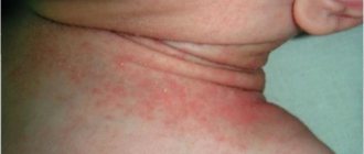
Peeling of the skin represents an accelerated process of necrosis of body surface cells with their exfoliation, which significantly worsens the appearance of the skin, disrupting the natural balance of tissue regeneration. Due to the influence of many reasons, such changes are observed, contributing to excessive drying of the surface and active rejection of epithelial cells.
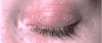
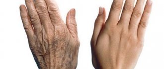
The visual characteristics of peeling represent the presence of small scales of the epidermis in certain areas of the skin, which manifests itself regardless of the type of skin, be it oily or combination. But most often people with dry skin suffer from such symptoms, when the root cause of peeling is genetically determined.
Make an appointment with a dermatologist by phone or by filling out the online form
| Select a clinic | Rash on back | Rash on cheeks | Rash on elbows |
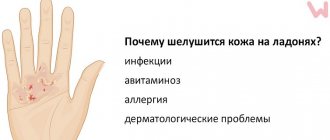
Why does skin peeling occur?
A disruption in the natural balance of renewal of the cellular structures of the skin can be observed for a number of reasons, the impact of which provokes a pathological deviation. Among them, the following should be highlighted:
- changes in ambient temperature;
- manifestation of an allergic reaction to the external use of care products;
- existing skin diseases;
- unbalanced diet, which is associated with the lack of fat-soluble vitamins necessary for the skin;
- natural aging of the surface layer;
- hereditary predisposition.
Thus, the causal effect contributes to a change in the ratio of the rate of keratinization of epithelial cells with their rejection, which leads to the manifestation of typical signs of skin peeling:
- itching;
- change in skin turgor;
- the appearance of a hint of hyperemia;
- presence of relative pain syndrome;
- formation of small cracks on the surface.
Transient jaundice
Yellow coloration of the skin and sclera with physiological jaundice does not appear from birth, but on the 3-4th day, reaching a maximum on the 5-6th day. The physiological process of destruction of red blood cells and the release of indirect bilirubin from them, as well as the immaturity of the liver enzyme system, leads to the accumulation of indirect bilirubin in the blood and skin, giving the latter a jaundiced tint. Sometimes transient jaundice has a protracted course. In this case, it is necessary to observe a pediatrician with an examination to exclude pathological conditions.
The principle of combating the manifestations of peeling
In certain cases, the appearance of pathological detachment of the epidermal layer develops against the background of the following diseases, which require specific treatment to eliminate the cause of the development of the pathology, and as a result, peeling of the skin disappears:
- seborrheic skin lesions;
- psoriasis;
- systemic lupus erythematosus;
- ichthyosis;
- dry erythema;
- ringworm and its varieties;
- scarlet fever;
- skin damage due to fungal infection and other conditions.
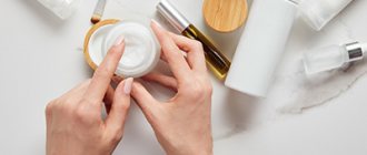
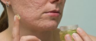
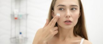
The development of pathology can be stopped on your own by using appropriate products for local application, maximally nourishing and moisturizing areas of the skin that are subject to the process of active exfoliation. In case of nutritional disorders, additional intake of fat-soluble vitamins helps restore the natural balance of cell layer renewal.
But if the condition worsens or the cause of the signs of pathology is unclear, you should consult a dermatologist who will competently conduct a diagnostic examination, which will help determine the most appropriate course of treatment. The main goal of diagnostics in this case is to exclude the presence of an infectious pathogen of any kind, which requires the subsequent use of specific treatment.
Useful information on the topic:
- Calling a dermatologist to your home
- Fungal infections
- Rash on the body
- Sexually transmitted diseases
- How to get tested
- Wart removal
- Removal of condylomas
- Tests for STIs
- Removal of papillomas
- Skin rash
- Pyoderma
- Skin structure
- Mushroom tests
- Diagnosis of sexually transmitted infections
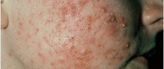
What diseases cause skin peeling?
There are many reasons that cause peeling skin. These include both ordinary allergies and more complex skin diseases. Dermatological diseases can be infectious or non-infectious. These include:
- Hereditary disease ichthyosis;
- Diseases of internal organs;
- Psoriasis;
- Dermatitis manifested in seborrhea;
- Lupus erythematosus;
- Fungal diseases of an infectious nature;
- Syphilis in secondary manifestations;
- Pityriasis rosea;
- Scarlet fever;
- Dry erytherma;
- Mycosis and xerosis.
With ichthyosis, the skin has a characteristic grayish tint. Its surface is covered with small scales. The patient experiences discomfort. His skin is peeling and itching. For this disease, no chemicals should be used. Even regular soap causes redness and irritation.
The main causes are sunburn, infections, HIV problems, dry skin in psoriasis, and skin injuries. Medicines play an important role. Seborrheic dermatitis occurs as a consequence of changes in the functioning of the skin. Sebum will be released in large quantities. This leads to progression of the disease.
Pityriasis rosea appears due to low levels of the body's immunity. This may be hypothermia of the dermis, insufficient nutrients. A cold or flu also worsens the condition of the body. It begins to react sensitively to external irritants in the form of soap, lotions, and other cosmetics. Cold wind, frost or hot weather increase pain.
Secondary syphilis appears as a skin rash. These invisible spots cause general malaise. The patient has a headache and a fever. Lymph nodes become larger. Peeling occurs suddenly. The deep layers need moisture. No amount of cosmetics can correct the situation. Treatment measures will give results if we find out the exact cause.
What tests need to be taken for peeling skin?
Deterioration of the condition of the skin can occur as a result of the use of bad cosmetics. In this case, it is enough to change your skin care system. But if the reasons are deep-rooted, then you must undergo a series of tests:
- General blood test. Necessary for identifying problems of an infectious nature, vitamin deficiency and anemia. This type of diagnosis allows you to identify helminths. They have a toxic effect on the body. This leads to deterioration of the dermis;
- Complete (biochemical) blood test. Its results make it possible to identify disturbances in the functioning of internal organs. You can find out what elements the body lacks for full development. For example, vitamins A and B affect skin condition. Their lack leads to flaking and dryness. Poor functioning of the kidneys, liver, stomach and all parts of the gastrointestinal tract also negatively affects our face, chin and other areas of the body.
- Blood test for general immunity level and determination of allergic reactions. A good allergy test can accurately find a specific allergen;
- Mycological examination. Prescribed to identify fungal infections in seborrheic dermatitis and pityriasis rosea;
- Histological examination. A scraping of the skin is taken in advance.
If a patient is suspected of having hormonal disorders, the doctor will refer you for a consultation with an endocrinologist. This specialist will determine the lack of hormones by ordering tests to determine thyroid hormones, estrogens (in women), and DHEA. With such an extensive examination, all causative factors can be determined. Draw up the correct treatment regimen. Develop a number of preventive recommendations.
Sexual crisis
This condition is observed in 2/3 of babies. Most often girls experience this. Rarely – premature babies. This phenomenon is associated with the reaction of the newborn’s body to release from maternal estrogens.
Often in the first week of life in girls, and sometimes in boys, the mammary glands slightly enlarge, but no inflammatory changes are observed on the skin. As a rule, engorgement increases by the 8-10th day of life, and by the end of the first month it subsides. Sometimes a fluid resembling colostrum is released from the baby's mammary glands. This phenomenon is normal! Parents should not be afraid.
In most cases, all of the above treatment does not require. BUT! If the engorgement of the mammary glands becomes significant, and the skin over them is red and hot to the touch, the body temperature rises and the child behaves restlessly, then a consultation with a pediatrician is necessary.
Do not squeeze liquid from your baby's nipples! This can cause pain to your baby and lead to infection.
In the first days of life, girls often experience grayish-white mucous discharge (possibly mixed with blood) from the external genitalia. These are manifestations of transient vulvovaginitis. After a few days, the discharge disappears. All this is also a normal reaction and does not require treatment. Hygienic care and regular washing are required.
What diet is necessary for peeling skin?
Balanced hypoallergenic diets will help fight the disease. First of all, you need to identify those products that cause painful reactions. Eliminate chocolate candies and bars from your daily diet. Citrus fruits, raspberries, strawberries, pomegranate, melon, black currants can also cause dryness. Honey, fish, nuts, mushrooms, cocoa, caviar are included in the list of such products.
Salads seasoned with vegetable oil have a healing effect. You should include porridge, eggs, dairy products, carrots, tomatoes, liver, and seeds. These products help increase the level of vitamins A and E. The lack of vitamin B can be compensated by unrefined cereals, green vegetables, avocados, bananas, carrots, walnuts, and cod fish liver.
Peeling of the skin due to gastrointestinal diseases
Painful signs associated with disturbances in the functioning of the gastrointestinal tract are immediately reflected in the condition of the skin:
- Gastritis and stomach ulcers cause acne, unnatural skin color, acne scars, greasy or dry skin;
- Diabetes, pancreatitis lead to itching, dryness, allergic reactions;
- Hepatitis and liver pathologies become the causes of the formation of age spots, jaundice, pimples, blackheads, asterisks, furunculosis;
- Gallbladder diseases contribute to the development of pimples, blackheads, jaundice, age spots;
- Duodenal ulcers, dysfunctions of the small intestine manifest themselves in unhealthy complexion, pimples, acne, allergic disorders, sagging, dryness;
- Constipation, diarrhea, disturbances in the functioning of the large intestine lead to fungal infections, sagging, dryness, acne, pimples and allergic spots.
If painful changes are detected, you should reconsider your diet. Consult a doctor for help, create an individual digestive program.
Common symptoms and manipulations in dermatology:
- Skin rashes
- Calling a dermatologist to your home
- Itching in the urethra
- Itchy skin
- Skin rash
- Prevention of casual sex
- Skin neoplasms
- Pyoderma
- Pityriasis rosea
- Streptoderma
- Scabies
- Peeling skin
- Fungal infections
- Skin infection
- Pus on the skin
- Blisters on the skin
- Papillomas on the foreskin
- Sexually transmitted diseases
- Skin structure
Mycoses of smooth skin
Among the widespread fungal diseases today, the most common are mycoses of smooth skin, such as microsporia, trichophytosis, lichen versicolor, mycosis of the feet (hands), and candidiasis. Sources of infection can be sick animals (cats, dogs, mouse-like rodents, cattle, etc.), as well as humans. In recent years, there has been an increase in the number of diseases caused by opportunistic fungi, among which superficial forms of candidiasis are most often recorded. Such a wide prevalence of these mycoses can be explained by the massive use of modern therapeutic agents, the environmental situation and other factors that reduce the body's defenses. One of the reasons for the significant prevalence of mycoses is the weakening of sanitary educational work in recent years. Due to insufficient awareness about the sources and ways of spreading infection, as well as adequate preventive measures, patients turn to the doctor late, and therefore mycoses become chronic, including in children suffering from mycoses of the scalp and smooth skin.
Microsporia is a fungal disease caused by various types of fungi of the genus Microsporum. In Russia, microsporia, which has spread over the last 50 years, is caused by a zoophilic fungus - fluffy microsporum (Microsporum canis), which parasitizes the skin of cats, dogs, and less often other animals. Infection from a sick person is observed in 2% of cases.
Epidemiology . Infection in 80-85% of cases occurs as a result of direct contact with a sick animal or through objects contaminated with the fur of these animals. Infection of children can also occur after playing in the sandbox, since the causative agent of microsporia is highly resistant to environmental factors and can remain viable in infected scales and hair for up to 7-10 years. Children often suffer from microsporia.
Clinic . After 5-7 days from the moment of infection, lesions appear on smooth skin, which can be observed on both open and closed parts of the body (children love to pick up animals and put them in bed with them). The lesions are round or oval in shape, pink or red, with clear boundaries, a raised ridge along the periphery, covered with blisters and thin crusts, with peeling in the center. The lesions are usually small, from 1 to 2 cm in diameter, single or multiple, sometimes merging. In 85-90% of patients, vellus hair is affected.
Treatment . If there are single foci of microsporia on smooth skin without damage to vellus hair, you can limit yourself to only external antifungal agents. The lesions should be lubricated with alcohol tincture of iodine (2-5%) in the morning, and sulfur-salicylic ointment (10% and 3%, respectively) should be rubbed in in the evening. You can rub the following antimycotics 2 times a day: mycozolon, mycoseptin, travogen or 1 time a day in the evening - mifungar cream, mycospor - until the clinical manifestations resolve. In case of multiple lesions of smooth skin and single lesions (up to 3) involving vellus hair, it is recommended to prescribe the antifungal antibiotic griseofulvin at the rate of 22 mg per 1 kg of the child’s body weight, in 3 doses after meals, in combination with exfoliating the stratum corneum of the epidermis in keratolytic lesions means (salicylic acid 3.0, lactic or benzoic acid 3.0, collodion up to 30.0). The lesions are lubricated with one of these products 2 times a day for 3–4 days, then 2% salicylic ointment is applied under compress paper for 24 hours, the detached scales of the stratum corneum of the epidermis are removed with tweezers and vellus hair is epilated. If during a control study carried out using a fluorescent lamp or microscope, affected hair is detected, the procedure is repeated. Detachment of the stratum corneum of the epidermis and manual hair removal of vellus hair can be carried out after using the “sealing” method. The lesions are sealed in a tile-like manner with strips of adhesive plaster for 2-3 days, this causes an aggravation of the process, which, in turn, facilitates hair removal.
The results of treatment for smooth skin microsporia are monitored using a fluorescent lamp or microscopic examination for fungi. The first control study is done after the resolution of clinical manifestations, then 3-4 days before the first negative test, and then after 3 days. The criteria for cure are resolution of the lesions, absence of luminescence and three negative tests on microscopic examination.
During the treatment, bed and underwear are disinfected: boiling in a soap-soda solution (1%) for 15 minutes (10 g of laundry soap and 10 g of caustic soda per 1 liter of water); ironing outerwear, furniture covers, and bedding five times with a hot iron through damp cloth.
Prevention. The main measure to prevent microsporia is compliance with sanitary and hygienic rules (you cannot use other people’s underwear, clothes, etc.; after playing with animals, you must wash your hands).
Trichophytosis is a fungal disease caused by various types of fungi of the genus Trichophyton. Trichophytons can be anthropophilic, parasitic on humans, and zoophilic, whose carriers are animals. Anthropophilic trichophytons include Trichophyton (Tr.) tonsuraus and Tr. violaceum, to zoophiles - Tr. mentagrophytes var gypseum and Tr. verrucosum.
Epidemiology. With superficial trichophytosis, caused by anthropophilic fungi, infection occurs through close contact with a sick person or indirectly through household items. Often children become infected from their mother, grandchildren from grandmothers suffering from a chronic form of the disease. The incubation period lasts up to a week. In zooanthroponotic trichophytosis, the sources of infection are sick animals: cattle, rodents. The highest incidence of this type of trichophytosis is recorded in the fall, which is associated with field work: it is at this time that the likelihood of infection through hay and straw increases. The incubation period ranges from 1–2 weeks to 2 months.
Clinic. On smooth skin with superficial trichophytosis, lesions can appear on any part of the skin - face, neck, chest, forearms. They have clear boundaries of a round or oval shape, with a raised ridge along the periphery of a bright red color; they are larger in size than in microsporia. The lesions are reddish-bluish in color, with peeling, nodules on the surface; in the chronic form, they develop on the skin of the buttocks, knee joints, forearms, less often the back of the hands and other parts of the body; the lesions do not have clear boundaries. Lamellar peeling is observed on the skin of the palms and soles. Vellus hairs are often affected.
With trichophytosis, caused by zoophilic fungi, the disease on the skin can occur in three forms: superficial, infiltrative and suppurative. Lesions are usually located on open areas of the skin. With a superficial form, they are round or oval in shape, with clear boundaries, a raised ridge along the periphery, on which bubbles, crusts, a pink center, and a bright red ridge are visible. The lesions are larger in size than with microsporia. Sometimes they are located around natural openings - eyes, mouth, nose. In the infiltrative form, the lesions rise above the skin level and are accompanied by inflammatory phenomena - infiltration. The suppurative form is characterized by the development of tumor-like formations, bright red in color, covered with purulent crusts due to the addition of a bacterial infection. When the lesion is compressed, pus is released from the hair follicles and pain is noted. The disease is accompanied by a violation of the general condition, sometimes the temperature rises. After the resolution of clinical manifestations, cicatricial atrophy of the skin remains at the site of former lesions. Clinical forms of zooanthroponotic trichophytosis can transform into one another.
Diagnostics. The diagnosis of trichophytosis is established on the basis of the clinic and when the fungus is detected during microscopy of pathological material, and the type of pathogen is determined using cultural examination.
Treatment. Treatment is carried out with antimycotics for external use. The lesions are lubricated with tincture of iodine (2-5%) during the day, and sulfur-salicylic ointment (10% and 3%, respectively) or mycoseptin is rubbed in in the evening. You can carry out monotherapy with ointment or cream (kanison, mifungar, mycozoral, mycospor (bifosin), exoderil, mycozoral, etc. In the infiltrative form, 10% sulfur-tar ointment is prescribed to resolve infiltration 2 times a day. Treatment of the suppurative form of trichophytosis begins with removing crusts from the affected area using bandages with 2% salicylic ointment, which are applied for several hours. After removing the crusts, vellus hair is epilated. Then apply lotions with solutions that have a disinfectant and anti-inflammatory effect (furacilin 1:5000, rivanol 1:1000 , potassium permanganate 1:6000, ichthyol solution (10%), etc.). As a result of this treatment, the hair follicles are freed from pus, inflammatory phenomena are reduced. Next, sulfur-tar ointment (5-10%) is prescribed for resorption of the infiltrate (5-10%) in the form of rubbing or under wax paper.After the infiltrate has resolved, antimycotics are used for external use (see superficial form of trichophytosis).In cases where vellus hair is affected in lesions on smooth skin, the stratum corneum of the epidermis is detached, followed by hair removal. To do this, you can use salicylic collodion (10-15%), milky-salicylic-resorcinol collodion (15%). If there is no effect, griseofulvin is prescribed orally at a daily dose of 18 mg per 1 kg of body weight, in 3 divided doses after meals daily until a negative test for fungi, then every other day. As an alternative method, terbinafine (Lamisil, Exifin) can be prescribed to adults 250 mg (1 tablet) once a day after meals every day, children weighing up to 20 kg - 62.5 mg, from 20 to 40 kg - 125 mg , over 40 kg - 250 mg in combination with antimycotics for external use.
The criteria for cure for trichophytosis are resolution of clinical manifestations and three negative fungal test results at three-day intervals.
Prevention. Prevention of trichophytosis depends on the type of pathogen. With superficial trichophytosis caused by anthropophilic fungi, the main preventive measure is to identify the source of infection, and it can be children suffering from superficial trichophytosis, or adults suffering from a chronic form of the lesion. In recent years, cases of chronic trichophytosis have been observed in middle-aged and older children. For suppurative trichophytosis, preventive measures are carried out jointly by medical workers, epidemiologists and veterinary services.
Mycosis of the smooth skin of the feet (hands). In a number of countries, up to 50% of the population suffers from mycosis of the feet. This disease is more common in adults, but in recent years it has often been observed in children, even infants.
Etiology. The main causative agents of mycosis of the feet are the fungus Trichophyton rubrum (T. rubrum), which is isolated in almost 90% of cases, and T. mentagrophytes var. interdigitale (T. interdigitale). Damage to the interdigital folds, which may be caused by yeast-like fungi, is recorded in 2-5% of cases. The anthropophilic fungus Epidermophyton floccosum is rarely isolated in our country.
Epidemiology. Infection with mycosis of the feet can occur in the family through close contact with a patient or through household items, as well as in a bathhouse, sauna, gym, or when using someone else's shoes and clothes.
Pathogenesis. The penetration of fungi into the skin is facilitated by cracks and abrasions in the interdigital folds caused by sweating or dry skin, abrasion, poor drying after water procedures, narrowness of the interdigital folds, flat feet, etc.
Clinic. Clinical manifestations on the skin depend on the type of pathogen and the general condition of the patient. The T.rubrum fungus can cause damage to the skin of all interdigital folds, soles, palms, dorsum of the feet and hands, legs, thighs, inguinal-femoral, intergluteal folds, under the mammary glands and axillary area, trunk, face, and rarely the scalp. The process may involve vellus and long hair, nail plates of the feet and hands. When the skin of the feet is affected, there are 3 clinical forms: squamous, intertriginous, squamous-hyperkeratotic.
The squamous form is characterized by the presence of peeling on the skin of the interdigital folds, soles, and palms. It can be flour-shaped, ring-shaped, lamellar. In the area of the arches of the feet and palms, an increase in the skin pattern is observed.
The intertriginous form is the most common and is characterized by slight redness and peeling on the lateral contact surfaces of the fingers or maceration, the presence of erosions, superficial or deep cracks in all folds of the feet. This form can transform into dyshidrotic, in which vesicles or blisters form in the area of the arches, along the outer and inner edges of the feet and in the interdigital folds. Superficial blisters open with the formation of erosions, which can merge, resulting in the formation of lesions with clear boundaries and oozing. When a bacterial infection is attached, pustules, lymphadenitis and lymphangitis occur. In the dyshidrotic form of mycosis, secondary allergic rashes are observed on the lateral and palmar surfaces of the fingers, palms, forearms, and shins. Sometimes the disease becomes chronic with an exacerbation in spring and summer.
The squamous-hyperkeratotic form is characterized by the development of foci of hyperkeratosis against the background of peeling. The skin of the soles (palms) becomes reddish-bluish in color, and pityriasis-like peeling is noted in the skin grooves, which extends to the plantar and palmar surfaces of the fingers. Pronounced ring-shaped and lamellar peeling may be detected on the palms and soles. In some patients, it is insignificant due to frequent hand washing.
In children, lesions of smooth skin on the feet are characterized by fine-plate peeling on the inner surface of the terminal phalanges of the toes, usually 3 and 4, or there are superficial, less often deep cracks in the interdigital folds or under the toes, hyperemia and maceration. On the soles, the skin may not be changed or the skin pattern may be enhanced; sometimes ring-shaped peeling is observed. Subjectively, patients are bothered by itching. In children, more often than in adults, exudative forms of lesions occur with the formation of blisters and weeping eczema-like lesions. They appear not only on the feet, but also on the hands.
Rubrophytia of smooth skin of large folds and other areas of the skin is characterized by the development of lesions with clear boundaries, irregular outlines, with an intermittent ridge along the periphery, consisting of merging pink nodules, scales and crusts, with a bluish tint (the color is bluish-pink in the center) . On the extensor surface of the forearms and shins, rashes can be located in the form of open rings. Lesions with nodular and nodular elements are often observed. The disease sometimes occurs as an infiltrative-suppurative trichophytosis (more often in men when localized in the chin area and above the upper lip). Foci of rubrophytosis on smooth skin can resemble psoriasis, lupus erythematosus, eczema and other dermatoses.
The fungus T. interdigitale affects the skin of the 3rd and 4th interdigital folds, the upper third of the sole, the lateral surfaces of the foot and toes, and the arch of the foot. This mushroom has pronounced allergenic properties. With mycosis of the feet caused by T. interdigitale, the same clinical forms of damage are observed as with rubrophytosis, but the disease is often accompanied by more pronounced inflammatory phenomena. In the dyshidrotic, less often intertriginous form, large blisters may appear on the skin of the soles and fingers, along with small blisters; in the case of bacterial flora, with purulent contents. The foot becomes edematous, swollen, and pain appears when walking. The disease is accompanied by an increase in temperature, deterioration of health, development of allergic rashes on the skin of the upper and lower extremities, torso, face, enlargement of the inguinal lymph nodes; the clinical picture is similar to that observed with eczema.
Diagnosis. The diagnosis is established on the basis of clinical manifestations, detection of the fungus by microscopic examination of skin flakes and identification of the type of pathogen by cultural examination.
Treatment. Treatment of mycosis of the smooth skin of the feet and other localizations is carried out with antifungal agents for external use. For squamous and intertriginous forms of lesions on the feet and other areas of the skin, medications are used in the form of a cream, ointment, solution, spray; you can combine a cream or ointment with a solution, alternating their use. Currently, the following medications are used to treat this disease: exifin cream, mycozoral cream, nizoral cream, canizon cream and solution, mycozon cream, mycospor (bifosin) cream, mifungar cream, lamisil cream and spray, mycoterbin cream. These drugs are applied to cleansed and dried skin once a day, the average duration of treatment is no more than 2 weeks. Antimycotics such as travogen, ekalin, batrafen, mycoseptin, mycozolon are used 2 times a day until clinical manifestations resolve, then treatment is continued for another 1-2 weeks, but once a day - to prevent relapse. In nodular and nodular forms of rubrophytosis, after relieving acute inflammatory phenomena using one of these ointments, sulfur-tar ointment (5-10%) is prescribed in order to further resolve the clinical manifestations. For intertriginous and dyshidrotic forms (the presence of only small blisters) of mycosis of the feet, drugs with a combined effect are used, which, along with an antifungal agent, include a corticosteroid, for example mycozolon, travocort, or a corticosteroid and an antibacterial drug - triderm, pimafucort.
In case of acute inflammatory phenomena (wetting, the presence of blisters) and severe itching, treatment is carried out as for eczema: desensitizing agents (intravenous or intramuscular administration of calcium chloride solution (10%), sodium thiosulfate solution (30%), calcium gluconate solution (10%) or calcium pantothenate orally; antihistamines. For external medications, at the first stage of therapy, lotions are used (2% boric acid solution, potassium permanganate solution 1:6000, 0.5% resorcinol solution), 1-2% aqueous solutions of methylene blue or brilliant green, fucorcin. Then they switch to pastes - boron-naphthalan, ichthyol-naphthalan, ACD paste - F3 with naphthalan, if complicated by bacterial flora - lincomycin (2%). At the 2nd stage of treatment after resolution of acute inflammatory phenomena, the above-mentioned antimycotic agents are used.
A drug such as Triderm, which contains, in addition to an antimycotic (clotrimazole 1%), a broad-spectrum antibiotic (gentamicin sulfate 0.1%) and a corticosteroid (betamethasone dipropionate 0), can quickly and effectively eliminate the symptoms of inflammation and itching in the presence of both fungal and bacterial infections. .05%). The presence of Triderm in 2 dosage forms - ointment and cream - makes it possible to use it for different types and at different stages of the pathological process.
If external therapy is ineffective, systemic antimycotics are prescribed: itraconazole in a continuous regimen of 200 mg per day for 7 days, then 100 mg for 1-2 weeks; terbinafine (Lamisil, Exifin) 250 mg once a day every day for 3-4 weeks; fluconazole (150 mg once a week for at least 4 weeks).
Prevention. To prevent foot mycosis, it is necessary to observe, first of all, the rules of personal hygiene in the family, as well as when visiting a bathhouse, sauna, swimming pool, gym, etc.; disinfect shoes (gloves) and linen during the treatment period. After visiting a bathhouse, swimming pool, sauna, to prevent mycosis of the feet, apply Daktarin spray powder to the skin of the interdigital folds and soles.
Tinea versicolor is a fungal disease caused by Malassezia furfur (Pityrosporum orbiculare), a yeast fungus. Lichen versicolor is quite widespread in all countries; young and middle-aged people suffer from it.
Etiology. Malassezia furfur as a saprophyte is found on human skin and, under favorable conditions, causes clinical manifestations.
Pathogenesis. Factors contributing to the development of the disease have not yet been precisely established, however, lichen versicolor is more common in people suffering from excessive sweating, changes in the chemical composition of sweat, diseases of the gastrointestinal tract, endocrine pathology, vegetative-vascular disorders, as well as immune deficiency .
Clinic. The disease is characterized by the presence of small spots on the skin of the chest, neck, back, abdomen, less often the upper and lower extremities, axillary and inguinal-femoral areas, on the head; the spots are initially pink in color and then become light and dark brown; Slight peeling is also observed, sometimes it can be hidden and can only be revealed by scraping. The rashes often merge, forming large areas of damage. After tanning, as a rule, white spots remain as a result of increased flaking. The disease is characterized by a long course with frequent exacerbations.
Diagnosis. The diagnosis is made on the basis of clinical manifestations, when the pathogen is detected in skin flakes during a microscopic examination and in the presence of a characteristic yellow or brown glow under a Wood's fluorescent lamp, as well as a positive iodine test.
Treatment. Currently, there is a sufficient selection of antifungal drugs for topical use that have a pronounced antifungal effect against the causative agent of lichen versicolor. These include imidazole and triazole derivatives, allylamine compounds. During the treatment of the disease, the following is used: exifin cream (applied to cleansed and dried skin in the affected areas 2 times a day for 7-14 days, if necessary, after a 2-week break, the course of treatment can be repeated), nizoral cream, mycozoral ointment, cream and canizon solution, mycozon cream, mifungar cream (prescribed once a day, duration of treatment is 2-3 weeks); lamisil cream and spray; nizoral shampoo (for three days, apply to the affected areas of the skin for 3-5 minutes and wash off in the shower). For common, often recurrent forms of lichen versicolor, systemic antimycotics are more effective: itraconazole (prescribed 100 mg once a day for two weeks, then take a two-week break, repeat the course of treatment if necessary), fluconazole (150 mg once a week within 4-8 weeks). During treatment, it is necessary to disinfect the patient’s clothes, hats, underwear and bed linen by boiling in a 2% soap-soda solution and ironing with a hot iron while wet. Family members of the patient should also be examined.
Prevention. To prevent recurrence of mycosis, it is necessary to use nizoral shampoo. Treatment should be carried out from March to May once a month for 3 days in a row.
Smooth skin candidiasis is a fungal disease caused by yeast-like fungi of the genus Candida.
Etiology. The pathogens are opportunistic fungi that are widely distributed in the environment. They can also be found on the skin and mucous membrane of the mouth, digestive tract, and genitals of a healthy person.
Epidemiology. Infection from the external environment can occur with constant fractional or massive infection with fungi.
Pathogenesis. Both endogenous and exogenous factors can contribute to the occurrence of candidiasis. Endogenous factors include endocrine disorders (usually diabetes mellitus), immune deficiency, severe somatic diseases and a number of others. The development of the disease is possible after the use of a number of modern medications: broad-spectrum antibiotics, immunosuppressive and hormonal drugs. The occurrence of candidiasis in the interdigital folds of the hands is facilitated by frequent contact with water, as this develops maceration of the skin, which is a favorable environment for the introduction of the pathogen from the external environment.
Clinic. On smooth skin, small folds on the hands and feet are more often affected, less often - large ones (inguinal-femoral, axillary, under the mammary glands, intergluteal). Lesions outside the folds are located mainly in patients suffering from diabetes mellitus, severe general diseases, and in infants.
In some patients, the disease begins in small folds of the skin with the formation of small, barely noticeable blisters on the lateral contacting surfaces of hyperemic skin, the process gradually spreads to the area of the fold, then peeling, maceration appears, or immediately shiny eroded surfaces of a deep red color with clear boundaries appear, with peeling of the stratum corneum of the epidermis along the periphery. The 3rd and 4th interdigital folds on one or both hands are most often affected. The disease is accompanied by itching, burning, and sometimes pain. The course is chronic, with frequent relapses.
In large folds, the lesions are dark red in color, shiny, with a moist surface, with a strip of exfoliating stratum corneum of the epidermis, occupying a significant surface, having clear boundaries and irregular outlines. New small erosions appear around large foci. In children, the process of large folds can spread to the skin of the thighs, buttocks, abdomen, and torso. Painful cracks sometimes form deep in the folds.
Candidiasis of smooth skin outside the folds has a similar clinical picture.
Diagnosis. The diagnosis is made on the basis of a typical clinic when a fungus is detected in scrapings from skin flakes during a microscopic examination.
Treatment. Limited and sometimes widespread acute forms of smooth skin lesions, especially those that developed during therapy with antibacterial drugs, as a rule, are easily treated with local antimycotic agents in the form of a solution, cream, ointment and can resolve even without treatment after discontinuation of antibiotics.
For candidiasis of smooth skin of large folds with acute inflammatory phenomena, treatment should begin with the use of an aqueous solution of methylene blue or brilliant green (1-2%) in combination with indifferent powder and continue for 2-3 days, then antifungal drugs are used until clinical resolution manifestations.
Among the antimycotic agents for candidiasis of smooth skin, the following are used: Canison solution and cream, Mycoson cream, Mifungar cream, Candida cream and solution, Triderm ointment and cream, Pimafucort, Pimafucin, Travocort, Travogen, Nizoral cream, Mycozoral ointment, Ekalin.
For common skin processes and in case of ineffectiveness of local therapy, systemic antimycotics are prescribed: fluconazole (Diflucan, Forkan, Mycosist) - adults at a dose of 100-200 mg, children at a dose of 3-5 mg per kg of body weight, itraconazole (100-200 mg), nizoral (adults 200 mg, children weighing up to 30 kg - 100 mg, over 30 kg - 200 mg) once a day daily, as well as the polyene antibiotic natamycin (adults 100 mg 4 times a day, children 50 mg 2–4 times a day). The duration of treatment is 2–4 weeks.
Prevention. Prevention of smooth skin candidiasis in adults and children consists of preventing its development in people suffering from underlying diseases, as well as in people receiving long-term antibacterial, corticosteroid, and immunosuppressive therapy. To prevent the development of candida infection in children hospitalized in somatic departments and receiving broad-spectrum antibiotics, it is necessary to prescribe fluconazole at the rate of 3 mg per kg of body weight once a day, treatment is carried out during the entire main course of therapy. Patients with intestinal candidiasis are prescribed nystatin 2–4 million units per day or natamycin 50 mg for children and 100 mg for adults 2 times a day for 15 days.
Zh.V. Stepanova, Doctor of Medical Sciences, TsNIIKV
Note!
- In recent years, there has been an increase in the number of diseases caused by opportunistic fungi, among which superficial forms of candidiasis are most often recorded.
- Due to insufficient awareness about the sources and ways of spreading infection, as well as adequate preventive measures, patients turn to the doctor late, and therefore mycoses become chronic
- 50% of the population suffers from mycosis of the feet. Adults get sick more often. Recently, there has been an increase in incidence in children, even infants.
- Treatment of mycosis of the smooth skin of the feet and other localizations is carried out with antifungal agents for external use.
- If external therapy is ineffective, systemic antimycotics are prescribed.
Peeling skin in children
The thickness of the top layer of skin in children is less than in adults. And therefore the causes of dryness are much greater:
- Prolonged exposure to the sun;
- Cold wind currents;
- Negative effects of soaps, shampoos and other care products;
- Lack of vitamins;
- Constant nervous disorders and stress;
- Infectious problems.
Children often experience chamomile, lichen, fungal infections, dermatitis, and eczema. Cases of hereditary diseases cannot be excluded.
All this is a consequence of poor nutrition and hygiene procedures based on low-quality products. Only a specialist can identify the causative factors. Therefore, as soon as parents see characteristic changes in the condition of the skin, it is necessary to seek help from a dermatologist.
Symptoms
Seborrheic dermatitis is manifested by redness, peeling with “oily” scales with a certain localization of foci.
In children - isolated on the scalp (in the frontal area it is called the “baby cap” or “milk crust”), behind the ears and in the external auditory canal, on the face (in the area of the eyebrows, wings of the nose), on the back of the neck (along the edge of hair growth), in areas of skin friction (in folds), under diapers.
Sometimes seborrheic dermatitis in children is widespread. These are rashes in the form of spots with greasy crusts, merging into larger areas. In some cases it is accompanied by itching.
In adults, seborrheic dermatitis is presented in the scalp area, on the face: in the area of the eyebrows, behind the ears, inside the ears, on the wings of the nose and in the areas of the cheeks adjacent to the nose.
It can manifest itself as separate spots with peeling, total damage to the scalp, damage to the eyelids in the form of blepharitis, damage to the chest wall in the form of individual nodules and spots with crusts on the surface, inflammation of the hair follicles on the skin of the upper half of the body (ostiofolliculitis).
Peeling skin in pregnant women
Skin with a dry surface indicates a lack of water in the body of a pregnant woman. The sebaceous glands do not work as actively. The thyroid gland may produce less hormones. Which is also a reason to see a doctor.
Sometimes a woman does not pay attention to such problems. Such inattention can lead to allergic reactions. Which are transmitted from mother to unborn baby. You need to see a doctor. Get examined and follow all medical recommendations.
Dryness itself cannot adversely affect the development of the fetus. But it can bring a lot of problems to a pregnant woman. Unsightly stretch marks appear. Microcracks and dandruff form in the hair. The body is itchy and itchy. For effective treatment, you should stop taking hormonal medications. Remove cosmetics containing aggressive substances from hygiene procedures.
It is not recommended to cleanse the skin with compounds that contain alkalis. Drinking clean water can eliminate problems such as itching and flaking. When caring for your skin, be sure to use nourishing oils. Choose cosmetics that contain natural substances. This causes a softening, sunscreen effect. Use liquid soap.
Types of skin peeling
With skin disorders, different types of peeling may occur:
- Pityriasis;
- Lamellar;
- Large-plate.
The characteristic features of the first type are small scales. But they cover the entire surface, forming different shades. It all depends on the presence of certain disorders within the body. Silvery-white scales occur with psoriasis. Yellowish - a consequence of seborrhea. Dark ones are formed when the disease is ichthyosis.
Lamellar peeling is accompanied by the formation of larger scales. In the large-lamellar form, rejection of the stratum corneum in entire layers is observed.
Prevention for peeling skin
Preventive measures for this disease are the first steps. You should pay close attention to your diet. After washing in the bath or shower, you need to use cosmetics that soften the skin. In cold weather it is necessary to protect yourself from wind and frost.
If you cannot solve the problems yourself, then seek the help of a specialist.
Methods for diagnosing skin diseases:
- Diagnosis of skin diseases
- Diagnosis of skin diseases at home
- Diagnosis of allergic skin diseases
- Diagnosis of bacterial skin diseases
- Diagnosis of viral skin diseases
- Diagnosis of hair diseases
- Diagnosis of nail diseases
- Diagnosis of skin tumors
- Blisters on the skin
- Dermatoscopy
- Demodex tests
- Diagnosis of sexually transmitted infections
- Mushroom tests
- Skin scraping
Uric acid infarction
Despite the menacing name, this condition should not cause concern for parents. Outwardly, it manifests itself as follows: in the first week of life, the baby produces cloudy, brick-yellow urine. This is explained by metabolic disorders in the kidneys and the deposition of crystals of uric acid salts in them. As a rule, by the end of the first week these phenomena disappear. If the color of urine does not return to normal by the end of the second week of life, you should consult a pediatrician for advice.
We have listed the main phenomena when faced with which young parents begin to sound the alarm. We hope that the article will be useful and you will be able to fully enjoy communication with your baby without worrying about transient conditions and knowing that this is a temporary phenomenon. Be healthy!
Call a dermatologist at home for peeling skin
Calling a doctor to your home will solve many problems. The patient does not have to make an appointment. He will be able to invite a specialist at a convenient time. In a home environment, it will be easier to talk about your problems. A dermatologist will be able to find out all the reasons related to the patient’s lifestyle. Draw up a treatment regimen after identifying all causative factors. In a confidential conversation, it would be better to offer recommendations on the use of cosmetics.
Transient catarrh of the intestines
Transient intestinal catarrh (physiological dyspepsia of newborns, transient intestinal catarrh) and transient dysbiosis are transitional conditions that develop in ALL newborns.
On the 1st-2nd day of life, babies pass their first stool (meconium), which is a thick viscous mass from dark green to black. On days 3-4, transitional stool appears - heterogeneous in consistency and color (with lumps, mucus, green-yellow color). By the end of the first week, the stool is usually set in the form of a yellow mush.
Transient dysbiosis is associated with the inevitable colonization of the intestinal mucosa by bacteria after birth. In subsequent months, the presence of undigested lumps in the stool, and occasionally mucus, may also be the norm if the baby has gained good weight and is feeling well. This is explained by physiological lactose deficiency. Young mothers should discuss this issue with their pediatrician.
Benefits of a private medical appointment
The result of seeking help from a dermatologist at a private specialized institution is the provision of not only advisory assistance at the appointed time, but also a full range of examinations with further implementation of the necessary treatment procedures. This fact greatly facilitates the implementation of all actions and provides comfortable conditions for completing the prescribed procedures.
And our Help Desk for private clinics in Moscow “Your Doctor” will help you find the right medical center that is convenient for visiting, which contains all the required data, allowing you to immediately make an appointment at a suitable time or arrange a call to a dermatologist at home.
Publication date: 2020-01-07
Useful information on the topic:
- Diagnosis of skin diseases
- Treatment of skin diseases
- Dermatology - the science of skin diseases
- How to consult a dermatologist
- Prevention of skin diseases
- Dermatologist appointment
- Examination by a dermatologist
- Pediatric dermatologist
- Skin doctor
- Paid dermatologist
This article is posted for educational purposes only and does not constitute scientific material or professional medical advice.