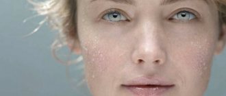
Rosacea is a common non-infectious chronic skin disease with a number of typical symptoms:
- Redness of the skin and the appearance of spider veins in the cheeks, cheekbones, nose and forehead;
- Itching and burning in the affected areas;
- Large rosacea - papules and pustules in the affected areas.
Around the world, about 10% of men and women of all ages suffer from the disease. Most often, rosacea manifests itself at the age of 30–40 years and begins with the expansion of small blood vessels in the skin of the face (less commonly, the chest and abdomen).
Not just acne
Rosacea or rosacea is a fairly common skin disease. The disease is characterized by persistent redness of the facial skin, spider veins, nodules and pustular pimples. Most often, rosacea occurs in people aged 30 years and older, and men get sick less often than women, but the course of the disease is more severe in the stronger sex. Rosacea begins with simple redness of the skin, which people do not pay attention to at first. Redness occurs as a reaction to frost, heat, hot and alcoholic drinks, spicy food or stress.
Etiology of rosacea
Experts identify a number of factors that initiate the development of acne. Most often this is severe overheating or, on the contrary, hypothermia of the skin, as well as:
- Exposure to sunlight, including in a solarium;
- Frequent stress;
- Local use of glucocorticosteroids;
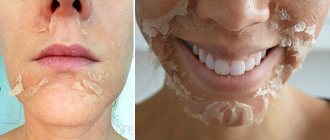
- The use of harsh scrubs not intended for facial skin;
- Independent use of chemical peeling;
- Bad habits: drinking alcohol and smoking;
- Constant inclusion of too hot and spicy foods in the diet.
If the patient has a history of individual intolerance to certain substances, manifested by allergic dermatitis, the risk of developing acne is quite high. It was previously thought that acne could be caused by demodex mites; recent research has found that this is not the case. Their presence in the follicles is not the cause of the development of the disease, but enhances its clinical manifestations.
Based on the method of exposure, it is customary to divide the factors for the development of acne into two groups:
- Exogenous - acting externally, represented by the physical effects of high and low temperatures, insolation, as well as nutritional - hot drinks, spices, etc.;
- Endogenous - gastrointestinal diseases caused by the bacterium Helicobacter pylori, infectious lesions of the skin, diseases of the endocrine system, decreased immunity.
Symptoms
Rosacea affects the skin of the face - cheeks, forehead, nose. Prolonged redness and spider veins appear, then small bright red nodules appear - papules, in the center of which purulent pimples appear. The rashes do not leave scars behind. A patient with rosacea feels itching and hot flashes to the face. The course of the disease is accompanied by frequent exacerbations and can last indefinitely. Over time, the skin of a patient with rosacea becomes rough and thick, and knobby formations appear. In some cases, lumpy hypertrophy of the skin of the nose develops - rhinophyma; deformation of the eyelids, ears, chin and forehead is also observed. There are no comedones with rosacea.
Etiology and pathogenesis
There is no single point of view about the origin and course of the disease, but according to the most common one, rosacea is a disorder associated with disorders of capillary innervation, primarily trigeminal nerve dysfunction. This is a mixed type cranial nerve - the largest of the twelve and is responsible for facial sensitivity, because. consists of very sensitive nerve fibers. Possible causes of the disease:
- exogenous climate forcing;
- poor nutrition;
- pathologies that have developed in the upper layer of the skin;
- microbe epidermal staphylococcus;
- mites of the genus Demodex (rosacea-like demodicosis);
- disruption of the gastrointestinal tract (for example, gastritis or ulcers);
- weak immunity;
- oxidative stress – cell damage;
- increased anxiety;
- insomnia;
- severe irritability;
- negative emotions;
- individuality of body constitution;
- exposure to proteins (cathelicidins, etc.) that protect the primary skin barrier from infectious pathogens that regulate blood pressure;
- hereditary factors;
- endocrine dysfunction, pregnancy, menopause (a relationship has been established between hemodynamic parameters in extracranial vessels and the content of individual hormones in the blood).
Dystonia and spasms play a negative role in the development of rosacea. Due to disruption of innervation, blood vessels become clogged. Externally, this is manifested by redness of the face and vascular networks on it. The facial vein supplies blood to the conjunctiva, the outer layer of the eyeball, which is why ophthalmic rosacea develops.
Modern dermatologists disagree about the etiology of rosacea. For example, the connection between the appearance of microorganisms on the surface of the skin and primary symptoms has not been clearly established. It has not yet been reliably established why the use of antibiotics always alleviates symptoms and improves the general health of the patient.
Stages of development
Dermatologists distinguish several stages of rosacea:
- the erythematous stage is characterized by dilated capillaries, spider veins, which occupy small symmetrical areas on the face, less often moving to the neck and chest;
- the papular stage is expressed by congestive redness, the presence of stars, superficial red or bluish nodules, hypertrophy of the sebaceous glands. Superficial pustules appear, which quickly disappear;
- The hypertrophic stage is characterized by persistent redness and bright red acne. These acne appear in groups, are usually follicular in nature, and last for several weeks. Gradually, a persistent papulopustular rash occupies the entire surface of the face, the skin of which becomes swollen and porous. The skin of the face thickens, acquires a bluish-purple color, becomes lumpy, and rhinophyma develops.
The lupoid or granulomatous form of the disease occupies a separate place and is characterized by the appearance of yellowish-brown or brownish papules, which give the “apple jelly” symptom on diascopy. According to the observations of our specialists, this stage of the disease is most often a consequence of treatment with corticosteroid ointments.
Atypical forms of rosacea
- Lupoid or granulomatous. Accompanied by the development of granulomas - yellow papules that leave scars on the skin after opening.
- Conglobat. Accompanied by large nodes, fistulas and the development of a purulent process.
- Halogen. Develops while taking bromine- or iodine-containing drugs.
- Steroid. Develops while taking steroid drugs.
- Gram negative. Develops against the background of uncontrolled or improper use of antibacterial drugs.
- Fulminant, or lightning fast. It most often occurs in young women and is characterized by the rapid development of all three stages of the disease. Accompanied by swelling and the formation of papules with purulent contents.
Treatment
Therapy for rosacea is a complex matter and largely depends on the stage of the disease. The course is selected individually. The doctor prescribes astringent and disinfectant lotions, pastes; electrocoagulation, dermabrasion, and cryotherapy work well. In particularly severe cases, antibiotics are prescribed. Vitamin therapy comes to the rescue in the treatment of rosacea. It is important to get rid of concomitant diseases: vegetative-vascular dystonia, gastrointestinal and hormonal disorders. Lifestyle requires complete correction. During treatment, it is important to follow a diet and eliminate provoking factors.
Phymatous form (rhinophyma) –
Phymatous changes are called thickening of the skin (mainly in the nose area) - with the appearance of roughness, irregularities, and nodules on it. The thickening can be pronounced and then it leads to partial disfigurement of the face. Occurs more often in men, because they are less likely to seek medical help in the early stages of the disease. Thickening of the skin of the nose is called rhinophyma. Rarely, the phymatous form can be observed on the chin, forehead, cheeks, ears and eyelids.
Also, with this form, telangiectasia and hyperplasia of the sebaceous glands can occur. In the dermal layer of the skin, a dense inflammatory infiltrate is observed, in place of which then, for example, tissue proliferation and fibrosis occurs with the formation of nodes. There are several histological types of rhinophyma, for example, fibrous, glandular, fibroangiomatous, etc.
Treatment regimens for the phymatous form –
| Expressiveness | Treatment |
| Mild course - an increase in skin thickness, but without changes in contours |
|
| Moderate flow - there is a change in contours, but without a nodal component | In addition to the above:
|
| Severe flow - there is a change in contours with a nodal component |
Rhinophyma: removal of excess tissue with a CO2 laser
Prevention
Rosacea is a vascular neurosis, which means that to prevent it, all provoking factors must be eliminated. Such factors include stress, heat and frost, wind and temperature changes, hot food and drinks, and alcohol. If occupational hazards have led to the development of rosacea (working in hot shops, staying at the stove for a long time, working in any weather in the field, on construction, at sea), you should change your profession or eliminate these hazards. When going outside, you need to protect your skin with special creams. Skin care products also need special ones designed for skin with rosacea.
Rosacea is not a contagious disease and is not life-threatening. However, rosacea, of course, does not bring joy, especially to women. So you shouldn’t start the disease if there is a chance to stop it at the very beginning.
Other articles by the author
- Exudative psoriasis
- Nail psoriasis
- Psoriasis of the palms and soles
- Red spots on the body
- Guttate psoriasis
- Plaque or vulgar psoriasis
- Rosacea
- Seborrhea. Acne vulgaris
- Seborrheic dermatitis
- Lichen planus
- Hives
- Psoriasis
Rosacea and related diseases
Introduction
The clinical and histological characteristics of rosacea remain poorly understood, despite the fact that rosacea is a relatively common dermatosis.
There is no laboratory test that can easily confirm the diagnosis of rosacea. Additionally, the idea that certain conditions are “related” to rosacea is based on clinical similarities rather than scientific evidence. The term “rosacea” covers a set of symptoms and signs that include persistent facial erythema, telangiectasias, inflammatory nodules and pustules, frequent facial flushes, non-pitting swelling of the face, inflammation of the eyes of various types, phymatous changes, mainly of the nose, as well as ears, forehead, chin, eyelids. Some authors distinguish between rosacea fulminans, characterized by the rapid appearance of nodules and pustules superimposed on facial erythema, sometimes with fever, and rosacea conglobata, when inflammatory cysts are observed on the face with subsequent scarring. However, the attribution of these symptom complexes to rosacea is controversial, and many authors consider them to be more related to acne vulgaris. Persistent red-brown nodules on the face with a characteristic granulomatous histology without caseating necrosis are called granulomatous rosacea, while frequent facial flushes and easily irritated facial skin are referred to as “prerosacea.” The inclusion of the above heterogeneous spectrum of clinical manifestations in the characteristics of rosacea makes it difficult to understand the pathogenesis of this disease [1]. In 2002, a classification of subtypes of rosacea was published, and it clarified the situation. Historical Background
Robert William is credited with the earliest medical descriptions of “acne rosacea.” The form of the disease that he described could nowadays be classified as papulopustular rosacea. Immediately, many dermatologists of that time began to believe that rosacea had a “seborrheic pathogenesis” similar to acne vulgaris. Radcliffe-Crocker later took it for granted that repeated hot flashes lead to dilated blood vessels in the face with subsequent inflammatory skin changes.
Epidemiology
Previously published reports regarding the incidence of rosacea in the population have sometimes been plagued by inappropriate definitions of the disease. In 1989, a study of more than 800 employees in Sweden found the prevalence of rosacea to be 10%. However, most of these individuals had facial erythema and telangiectasias without inflammatory skin lesions, signs that are clinically indistinguishable from facial skin changes caused by chronic exposure to ultraviolet light (dermatoheliosis). A demographic study of 1000 people in Ireland, which applied a consensus clinical definition of papulopustular rosacea, found the prevalence of rosacea to be 2.7%. This study showed that papulopustular rosacea is not associated with photodamage. An additional 14% of this study population had skin changes similar to those in the Swedish group, which could be attributed to either erythematotelangiectatic rosacea or dermatoheliosis, disorders associated with photodamage.
Pathogenesis
The exact pathogenesis of rosacea is unknown. It will likely be established over time that the disease now known as rosacea comprises several similar, possibly related, but distinct clinical conditions, each with its own predominant pathogenetic mechanism. Some factors that are relevant to rosacea are listed in Table 1.
Most often, rosacea is observed in people of the Caucasian race and in people with fair, sun-sensitive skin, i.e. with phototypes I and II. There may be a genetic predisposition to this condition, as 10−20% of patients report a family history of rosacea. Epidemiological studies suggest that erythematotelangiectatic rosacea may be associated with ultraviolet radiation exposure and photodamage. It has been established that exposure to ultraviolet B radiation stimulates angiogenesis and increases the secretion of VEGF by keratinocytes. It is assumed that exposure to UVR leads to the production of reactive oxygen, which, by causing increased production of matrix metalloproteinases, causes damage to blood vessels and the main intercellular substance of the dermis. It is possible that these factors are especially significant in patients with erythematotelangiectatic rosacea. Histological examination of papulopustular rosacea reveals inflammatory changes, which are most pronounced in the area of the pilosebaceous follicle. These changes are likely the result of dysfunction of the innate immune response, which protects the skin from infection as well as other environmental irritants such as UVB and chemical trauma. Triggering of the innate immune system results in the controlled secretion of cytokines and antimicrobial peptides such as cathelicidin. In rosacea, there is upregulation of cathelicidin and serine protease processing (kallekrein 5), indicating dysfunction of the innate immune system. When cathelicidin peptides and related enzymes were injected into the skin of mice, they stimulated pro-inflammatory and angiogenic activity, properties that may be of greater relevance to papulopustular rosacea. Persistent centrofacial erythema and telangiectasia are common signs of rosacea, as are hot flashes (transient erythema). Compared with unaffected skin, skin affected by papulopustular rosacea showed increased cutaneous blood flow. In addition, expression of VEGF, CD31 (a common endothelial cell marker), and the lymphatic endothelial marker D2-40 (podoplanin) is increased in skin lesions, suggesting stimulation of endothelial cells of blood and lymphatic vessels. Vasodilation is presumably explained by impaired control of skin thermoregulatory mechanisms. Compared to controls, patients with rosacea blush more easily in response to heat and have been shown to have a lower temperature pain threshold [1]. Patients with rosacea often report symptoms of burning, burning, and dryness on their face. Studies have confirmed a reduced skin irritability threshold in patients with rosacea. This may occur due to epidermal barrier dysfunction, in which transepidermal water loss increases in patients with rosacea. In addition, it is assumed that damage or pathology of the stratum corneum leads to the penetration of sensory stimuli, which causes a burning sensation. These findings have practical implications for counseling patients with rosacea regarding appropriate facial skin care. Neurogenic inflammation (an inflammatory response caused by sensory nerves that secrete neurotransmitters at the site of inflammation) can lead to vasodilation, extravasation of plasma proteins, and recruitment of inflammatory cells. It is known that environmental triggers activate G protein-coupled receptors (serpentines) and ion channels on sensory nerves; following stimulation, neuropeptides such as calcitonin gene-encoded peptide and adenylate cyclase-activating polypeptide cause vasodilation of arterioles and capillaries. However, these pathogenic pathways are not well understood and their relevance to rosacea is unclear. Although Demodex mites (folliculorum and brevis) are commensals of the facial skin, they are often observed in pilosebaceous follicles in histological examination of skin biopsies of patients with rosacea. Using superficial skin biopsy techniques, several studies have found that these mites are present in much higher numbers in patients with rosacea than in controls. It has been shown that the invasion of multiple mites into the follicles is associated with a pronounced perifollicular infiltrate, predominantly with CD4+ T helper cells. Antigenic proteins produced by a single bacterium (Bacillus oleronius) isolated from Demodex mites have been found to stimulate the inflammatory response in patients with papulopustular rosacea. It has been hypothesized that Demodex mites and associated bacteria upregulate skin proteases, thereby enhancing the dysregulation of the local innate immune response. It is likely that these pathogenetic mechanisms are very important for the papulopustular subtype of rosacea. Finally, given current evidence, it seems unlikely that Helicobacter pylori infection plays an etiological role in the pathogenesis of rosacea.
Clinical picture
Rosacea usually occurs in middle age, with women becoming affected relatively earlier than men. Rosacea has been reported in children with associated ocular changes and treatment responses that are similar to those in adult patients, but the lack of adequate criteria to confirm the diagnosis prevents the establishment of juvenile rosacea as a disease entity. Although there are reports that women are affected more often than men, there is no clear evidence of this. Rhinophyma, however, occurs mainly in men. The prevalence of rosacea among people with darker skin phototypes has not been studied in detail, but it may be greater than previously thought. In highly pigmented skin, erythema and telangiectasia are more difficult to assess clinically, in such cases a history of skin sensitivity and hot flashes, together with centrofacial or inflammatory ocular lesions, will aid in diagnosis. In 2002, a classification scheme for rosacea was presented (Table 2) and the clinical characteristics of its 4 main forms.
Rhythematotelangiectatic rosacea
diagnosed in people suffering from hot flashes against a background of persistent facial erythema and, sometimes, telangiectasia (Fig. 1) [2]. Such patients usually have skin phototype I or II. Clinical recognition of erythematotelangiectatic rosacea may be difficult due to overlap with skin signs of chronic sun damage in fair-skinned individuals (dermatoheliosis). In addition, patients with dermatoheliosis may have thermolabile facial skin and experience hot flashes, especially with changes in environmental temperature. Patients who complain of hot flashes as their only symptom should not be classified as having “prerosacea” and should rather be evaluated for other causes of hot flashes.
Papulopustular rosacea
characterized by the appearance of small inflammatory dome-shaped papules against the background of hyperemia and telangiectasias.
The most common location of papules is the central part of the face and its convex areas (Fig. 2) [2]. These lesions are found singly or in clusters. Patients complain of mild soreness or itching at the affected areas, but the social distress caused by the appearance of this rash far outweighs the physical symptoms. Individual lesions persist for about 2 weeks, then turn into macular erythema, which gradually disappears; no final scarring is observed. Disappeared lesions are replaced by new ones. A rim of erythema may surround larger inflammatory lesions, and small telangiectatic blood vessels are visible within this rim. Severely affected skin sometimes develops slight flaking or crusting. These signs should not be confused with seborrheic dermatitis, which can accompany papulopustular rosacea and is characterized by greasy, yellowish scales and involvement of the eyebrows and facial folds. Phymatous rosacea
is characterized by an increase in the thickness of the epidermis, knobby growths of the soft tissues of the nose, forehead, chin, ears, and cheeks. The types of phymatous rosacea are presented in Table 3. The most common manifestation of phymatous rosacea is rhinophyma, which is formed due to hypertrophy of the sebaceous glands in the skin of the nose (Fig. 3) [2]. Rhinophyma is usually seen in patients with other features of rosacea, but it can occur in patients with acne vulgaris; Sometimes rhinophyma is caused by chronic sun damage or occurs de novo. Rhinophyma predominantly affects men. It should be noted that rhinophyma does not represent the end stage of rosacea, since most patients had only mild or no rosacea. The earliest clinical sign of rhinophyma is the appearance of enlarged pores (open follicles) on the distal parts of the nose. In severe cases, there is tissue hypertrophy with nasal deformation. Important note: The edematous changes sometimes seen in patients with rosacea should not be confused with phymatous rosacea, as such changes (unlike changes in phymatous rosacea) often improve with treatment of the inflammation.
Ophthalmic rosacea
(Fig. 4) [2] may not be accompanied by skin changes (in which case it is difficult to establish a correct diagnosis) or may be present in patients with any other subtypes of rosacea. Apparently, patients with erythematotelangiectatic and papulopustular rosacea are especially susceptible to developing eye inflammation - up to 50% of such patients suffer from this pathology. Symptoms of ophthalmic rosacea: itching, lacrimation, dryness and a feeling of sand in the eyes, the appearance of crusts on the eyelids, frequent styes and, as a result, the inability to wear contact lenses. In other cases, patients may not present relevant complaints due to mild symptoms. Clinical signs of ophthalmic rosacea are diverse and lack specificity. There may be small deposits at the base of the eyelashes (conical scales) or moderate peeling of the edges of the eyelids. A more active form of the disease causes blepharitis, often with swelling of the eyelids and injection of the conjunctiva, the latter giving the affected eye a reddish tint. Cysts arising from the meibovial glands (chalazion) are firm, painless swellings on the outer surface of the tarsal cartilage, while hordeolum (styes) are a similar but painful inflammation. Fortunately, severe eye disease (keratitis, corneal neovascularization, uveitis, scleritis, or iritis) is rare in patients with rosacea. Patients who complain of pain or photophobia require an examination by an ophthalmologist [3].
In addition to the 4 main types of rosacea, there are variations that include granulomatous rosacea and rosacea fulminans, also called pyoderma faciale. Rarely, rosacea lesions are located outside the face, mainly on the scalp in patients with androgenetic alopecia.
Histopathology
Histological changes in the erythematotelangiectatic form (Fig. 5) of rosacea may be subtle and are often limited to vascular ectasia and slight edema. In the inflammatory papulopustular form (Fig. 6), a more pronounced perivascular and perifollicular lymphohistiocytic infiltrate appears. Some patients may have severe hyperplasia of the sebaceous glands. Comedones are not formed. In granulomatous rosacea (Fig. 7), epithelioid granulomas without curdled necrosis are visible in the dermis [4].
Differential diagnosis
The differential diagnosis of papulopustular rosacea is made with acne vulgaris. However, unlike acne vulgaris, open and closed comedones do not occur in patients with rosacea. Since both conditions are quite common, patients with rosacea may have signs of coexisting or preexisting acne vulgaris (eg, scarring). It is also necessary to take into account rosacea-like diseases, especially periorificial dermatitis and steroid-induced rosacea. The differential diagnosis also includes demodex folliculitis. The erythematotelangiectatic form of rosacea must be distinguished from chronic sun damage (in fair-skinned individuals), seborrheic dermatitis, tuberculous lupus (no pustules), lichen planus pilaris, and erythromelanosis of the face. The last two diseases have characteristic small white papules. The differential diagnosis of blepharitis in ocular rosacea includes seborrheic dermatitis and allergic contact dermatitis [5].
Treatment
The division of rosacea into subtypes helps in choosing therapy (Table 4). The identified subtypes determine the choice of specific treatment: thus, the treatment of acute inflammation and the necessary measures to maintain remission will differ. Depending on the degree - mild, moderate, severe (I, II, III) - one or another treatment option is prescribed. The classification is also used to monitor the course of the disease.
At the initial consultation, it is important to give the patient an understanding of the chronic, relapsing nature of his disease and the need for systematic maintenance treatment. Treatment includes facial skin care, suppressing the tendency to hot flashes by avoiding provoking factors and using special medications, and removing telangiectasia. It is important to avoid exposure to the sun and constantly use sunscreen (protection factor of at least 15), since a significant part of the patient's skin changes may be due to dermatoheliosis. Patients with papulopustular rosacea are primarily treated with topical and systemic antibiotics, used alone or in combination. Sometimes successful treatment of papulopustular rosacea makes the underlying telangiectasias visible. It is a good idea to warn patients about this possibility, otherwise they may attribute the appearance of these telangiectasias to the treatment being administered. After successful treatment of inflammatory lesions, patients with papulopustular rosacea should continue supportive therapy (usually local); without observing this rule within a period of 3 to 6 months. relapse is likely. Many patients with moderate to severe papulopustular rosacea (grades II and III) require repeated courses of systemic antibiotic therapy. Rhinophyma is the most common manifestation of phymatous rosacea and is the one most susceptible to surgical treatment. Mild disease may respond to drug treatment with isotretinoin, although convincing evidence of effectiveness is lacking. More severe disease (grades II and III), with noticeable enlargement and deformation, responds best to physical treatments, such as CO2 laser therapy, electrosurgery, or surgical excision. Other variants of phyma are very rare, and therapeutic interventions are based solely on anecdotal reports. Patients with mild ophthalmic rosacea (grade I) often complain of itching and a dry, gritty feeling in the eye(s). Daily eyelid hygiene and artificial tear replacement treatment are usually sufficient for this degree of ocular rosacea. Burning or sore eyes with crusting of the lid margins or the formation of chalazions and styes are moderate (grade II), and patients require topical antibiotics (eg, metronidazole gel or fusidic acid) or systemic antibiotic therapy, as for papulopustular rosacea. Symptoms such as pain or photophobia, as well as visual disturbances, are signs of severe illness (grade III). Such symptoms and lack of response to treatment require urgent referral to an ophthalmologist.
Rosacea-like diseases
Morbighan's disease is a rare disease characterized by progressive, asymptomatic, non-pitting swelling in the upper central part of the face, which is combined with fixed facial erythema (Fig. 8).
The exact etiology is unknown. Histological examination reveals edema of the dermis with predominant mast cells in a moderate inflammatory infiltrate. Morbighan's disease may be the result of various triggers, such as trauma or allergic contact dermatitis, and its association with rosacea is controversial. There is also a point of view that Morbighan disease is a complication of chronic rosacea, namely lymphedema of the upper half of the face and ears [1]. The conclusion is disappointing: there is no effective treatment for this pathology, although long-term courses of low-dose isotretinoin have been reported to help some patients and that systemic antihistamines have a similar effect. Jansen and Plewig [7] proposed a treatment regimen including isotretinoin at a dose of 0.1−0.2 mg/kg per day for 2−4 months, which can be combined with ketotifen at a dose of 1−2 mg per day and an antihistamine means. Periorificial dermatitis, also called perioral dermatitis (Figure 9), is similar in appearance to rosacea, but the morphology of the lesions (grouped superficial small papulovesicles or papulopustules at the same stage of development, rather than larger clusters of papules and pustules at different stages of development) , and perioral rather than centrofacial distribution usually indicate the correct diagnosis. In addition, papules may overlap or intersperse with thin plaques, which resemble nonspecific dermatitis. Sometimes papules have a granulomatous component. These patients may have a history of use of topical or inhaled corticosteroids. Affected people often describe intolerance to the sun, cosmetics and/or hot water. For such patients, systemic antibiotics (tetracycline, doxycycline, erythromycin, azithromycin) are prescribed for a period of 4 to 6 weeks. Local corticosteroid therapy should be discontinued, and the use of cosmetics should also be discontinued. Unlike rosacea, recovery is usually not accompanied by subsequent relapse. Steroid-induced rosacea
(Figure 10) presents with erythema, papules, pustules, sometimes telangiectasia, and atrophy due to repeated facial applications of potent topical corticosteroids. Patients suffering from steroid-induced rosacea are often unaware of the causal relationship between steroid application and the rash. On the contrary, the application of steroids usually causes a rapid, even short-term improvement in symptoms, creating the illusion of a pronounced beneficial effect.
Demodex mites are present in large numbers. Reducing the intensity of topical steroid applications or switching to a less active steroid, rather than abrupt withdrawal, will help avoid the rebound phenomenon. Complete clearing of the skin usually requires systemic antibiotic therapy (tetracycline or doxycycline) for up to 6 weeks. Haber syndrome
– a hereditary symptom complex characterized by a rosacea-like rash on the skin of the face, aggravated by exposure to sunlight, and small plaque-like areas of excessive keratinization of the skin on the trunk, histologically corresponding to intraepidermal epithelioma. The type of inheritance is autosomal dominant. The disease manifests itself in children as persistent erythema on the skin of the nose, cheeks, and chin. Gradually, persistent telangiectasias, small dense, red nodules, small areas of peeling, and mild atrophy form against its background. Small plaque-like (up to 1 cm in diameter) keratotic or scaly lesions form on the skin of the chest and back. The diagnosis is based on the clinical picture and histological findings. Differential diagnosis is carried out with red acne, photodermatitis, which are not characterized by intraepidermal epitheliomas.
Using the M22 platform provides:
- comprehensive solution to the problem, eliminating all clinical manifestations of rosacea, getting rid of pathological microorganisms and accelerating skin regeneration;
- an effect that will not keep you waiting. Visible improvements in skin condition are observed after the first procedure;
- safety and absence of side effects;
- no rehabilitation period;
- personalized approach to each client, thanks to the availability of a variety of attachments and choice of light emission characteristics.
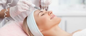
Stages of the procedure
At the initial consultation, the doctor examines the patient and draws up an individual treatment plan. Then the specialist selects the necessary attachment for the M22 device and the range of characteristics, taking into account the characteristics of the course of the disease. During the procedure, the patient must wear protective glasses. The doctor performs sequential treatment of the affected areas of the skin using light radiation. The procedure is absolutely bladeless and comfortable. The duration of sessions and their frequency depend on the complexity of the disease and its type.
Equipment
Fraxel re:store
A laser that has gained wide recognition in more than 80 countries around the world.
M22™ device
M22 is the standard of multifunctional lighting equipment for aesthetic cosmetology from Lumenis.
Antera 3D
Used for diagnostics before cosmetic procedures, as well as for subsequent assessment of their effectiveness.
Diagnostic methods
The diagnosis of rosacea is made by a dermatologist based on an external examination. To identify the causes of the disease and exclude pathologies, laboratory diagnostic methods are used. Depending on the form of the disease, the following methods can be used:
- scraping the skin to determine the presence of a skin mite;
- examination of the contents of papular elements;
- blood tests for markers of systemic diseases (especially lupus erythematosus).
Rosacea can be mistakenly confused with acne, so it is important to see an experienced professional.