What causes skin pigmentation disorders?
Bibliography
1 GRLS, r/u LP-004175 dated 03/03/2017
2 Certificate GMP-0036-000221/18
3 Greveling K, Prens EP, Ten Bosch N, van Doorn MB. Comparison of lidocaine/tetracaine cream and lidocaine/prilocaine cream for local anesthesia during laser treatment of acne keloidalis nuchae and tattoo removal: results of two randomized controlled trials. Br J Dermatol. 2016 Jul 5. doi:10.1111/bjd.14848 Hernandez E.;
4 J. Cassuto, R. Sinclair, M. Bonderovic, Anti-inflammatory properties of local anesthetics and their present and potential clinical implications. Acta Anaesthesiol Scand 2006; 50; 265-282]
5 Based on IQVIA report March 2022 - September 2022
6 Russian medical journal. Zhigultsova T.I., Parkaeva L.V., Ilyina E.E., Vissarionov V.A.: “Experience of using 5% Emla cream in the practice of dermatocosmetologists”
7 Instructions for use of the medicinal product for medical use Acriol Pro
8 T.N. Calvey, N.E. Williams. Pharmacology for anesthesiologists. Publishing house Binom, Moscow, 2007, 119-128
9 Based on the report “Anesthetics in injection cosmetology for 2016” Vademecum Analytical Center for sales volume.
10 T.I. Zhigultsova, Ph.D. L.V. Parkaeva, E.E. Ilyina, professor V.A. Vissarionov: “Experience of using 5% Emla cream in the practice of dermatocosmetologists” Cosmetology and plastic surgery. Vol. 16, No. 9, 2008
11 On the Russian market there are so-called cosmetic products containing lidocaine and not registered as medicines
12 Cling film can be used as an occlusive dressing
13 Drug Release Studies on an Oil-Water Emulsion Based on a Eutectic Mixture of Lidocaine and Prilocaine as the Dispersed Phase
14 Federal Law-61 “On the Circulation of Medicines” dated April 12, 2010, Federal Law-323 “On the Fundamentals of Protecting the Health of Citizens in the Russian Federation” dated November 21, 2011 and Federal Law-532 “On Amendments to Certain Legislative Acts of the Russian Federation Regarding Counteraction to Trafficking falsified, counterfeit, substandard and unregistered medicines, medical devices and counterfeit dietary supplements" dated December 31, 2014.
15 Federal Law-532, Technical Regulations of the Customs Union on the safety of perfumery and cosmetic products, Federal Law 61, Federal Law 532, Criminal Code of the Russian Federation, Art. 235, 238, 227
16 V.V. Osipova.MMA named after. I.M. Sechenov. Psychological aspects of pain. Lecture. №1/2010
17 According to GMP News. Analysis of the market for local anesthetics used in cosmetic injections in 2022.
18 One type of occlusive dressing, namely: bandage, cling film, adhesive tape or rubberized fabric
19 The combination of lidocaine and prilocaine in concentrations above 0.5-2% has bactericidal and antiviral properties. In Akriol Pro the concentration is 5%.
20 A.A. Stepanov, G.V. Yatsyk, L.S. Namazova Method of preventing pain in young children during vaccination // Into the practice of pediatricians - 09.14.2006 -
21 N.V. Klipinina, RMJ, Some features of the perception and experience of pain by children: a psychologist’s view, reprint. 2007.1-7
22 E.A. Ranneva. The use of EMLA® cream in the complex correction of cosmetic imperfections. Experimental and clinical dermatocosmetology 2010, No. 2: 48-53.
23 Gonzalez S. Evaluation of Topical Anesthetics by Laser-Induced Sensation. Lasers in Surgery and Medicine 23:167–171(1998));
24 V.G. Lebedyuk et al. Anesthesia in dermatocosmetology. Experimental and clinical dermatocosmetology, 2010 No. 5
25 Meltem F. Söyleva Nilüfer Koçaka Bahar Kuvakia Seyhan B. Özkanb Erkin Ki˙rb; Anesthesia with Cream for Botulinum A Toxin Injection into Eyelids. Ophthalmologica 2002;216:355–358
26 E.V. Matuszewska and authors. Topical local anesthetics in cosmetology. Clinical Dermatology and Venereology. 03.2017 pp. 89-96
27 Therapeutics and Clinical Risk Management 2006:2(1) 99 – 113
28 F. Michael Ferrante, Timothy R. Wade Bopcore Postoperative Pain. Management. Per. from English/Ed. M.: Medicine, 1998.- 640 pp., p. 243
29 Lakhin R.E. Local anesthetics. Department of Anesthesiology and Reanimatology of the Military Medical Academy named after. S.M.Kirova, St. Petersburg, 2013, Committee on Ultrasound Technologies of the All-Russian public organization “Federation of Anesthesiologists and Resuscitators.” Clinical guidelines “Intensive therapy for systemic toxicity with local anesthetics. Moscow - St. Petersburg 2015 Page. 10 https://www.far.org.ru/recomendation
30 Wetter DA et al. J American Acad. Dermatol. 2010;63(5):789-98
31 https://www.1nep.ru/articles/issledovanie-sostava-populyarnykh-mestnykh-anestetikov/
32 ORDER OF THE MINISTRY OF HEALTH OF THE RUSSIA dated 08.10.2015 No. 707n
33 Davydov O.S. Peripheral and central mechanisms of the transition of acute pain to chronic pain and the possible role of cyclooxygenase 2 inhibition in the prevention of chronic pain syndrome. Neurology, neuropsychiatry, psychosomatics. 2016;8(2):10-16.
34 J. ALASTAIR CARRUTHERS, MD, JEAN DA CARRUTHERS, MD. Safety of Lidocaine 15% and Prilocaine 5% Topical Ointment Used as Local Anesthesia for Intense Pulsed Light Treatment. Dermatologic Surgery 2010;36:1130–1137
35 Ya-Xian et al. The number of cells in the stratum corneum of normal skin depending on the anatomical location on the body, age, gender and physical parameters Archives Dermatol Res 1999; 291:555–559.
36 J. Morgan, Magid S. Michael. Clinical anesthesiology, Book 1. Binomial. Moscow St. Petersburg, 2001
37 Arendt-Nielsen L, Bjerring P, Nielsen J. Acta Derm Venereol 1990;70:314-318
38 Belarusian State Medical University. 2nd Department of Therapeutic Dentistry. Therapeutic dentistry. Part 1. Ed. A.G. Tretyakovich, L.G. Borisenko.
39 Dermatol Surg 1999;25:950-954
40 K. Greveling et al. Br J Dermatol 176(1), 81-86. 2016 Dec 10
41 Juhlin and Evers Adv Dermatol 1990;5:75-92
42 Arendt-Nielsen L, Bjerring P, Nielsen J. Acta Derm Venereol 1990;70:314-318
43 Study Desensor 001. Wahlgren CF, Quiding H. J Am Acad Dermatol 2000;42:584-8.
44 https://www.1nep.ru/estetic/articles/13952/
45 Guide to dermatocosmetology, edited by E.R. Arabia and E.V. Sokolovsky. St. Petersburg: Foliant Publishing House LLC, 2008 - 632 pp.
46 https://www.1nep.ru/estetic/articles/132197/
47 https://medside.ru/dikain
48 https://medi.ru/instrukciya/novokain_9473/
49 https://medside.ru/anestezin
50 https://grls.rosminzdrav.ru/Default.aspx
51 Radman et al.2002, Yamashita., 2003
52 https://www.krasotaimedicina.ru/diseases/hematologic/methemoglobinemia
53 Evers H, Scott B, Dahlquist AC. Dermal analgesia after epicutaneous application of 5% cream, 5% prilocaine cream, 5% lidocaine cream and placebo cream, to volunteers. Study 89EM03 (n= 21, cross-over). CSR 802-10AC088-2,1989.
54 MASMI. A study of users of skin anesthetics. June 2018
55 O.M. Burylina, A.V. Karpova. Cosmetology. Clinical guidelines. GEOTAR-Media. Moscow, 2018
56 Postoperative pain: the role of peripheral and central sensitization mechanisms. https://rsra.rusanesth.com/publ/posleoperatcionnaya_bol.html
57 Paul M. Friedman, MD, Jushua P. Fogelman, MD and others. Comparative Study of the Efficacy of Four Topical Anesthetics. Dermatolog Surg 1999; 25:950-954
58 https://www.1nep.ru/articles/rynok-kosmeticheskikh-anestetikov-dlya-kozhi/
59 https://www.rlsnet.ru/mnn_index_id_879.htm
60 https://yandex.ru/health/pills/product/ultrakain-ds-2195
61 https://grls.rosminzdrav.ru/Default.aspx on 02/15/2021
62 Ziganshin O.R. Comparison of the effectiveness and safety of topical local anesthetics for superficial surgical interventions in dermatology. Clinical dermatology and venereology. 2018;17(6):53-60. https://doi.org/10.17116/klinderma 20181706153
63 Comparison of Topical Anesthetics for Radiofrequency Ablation of Achrocordons: Eutectic Mixture of Lignocaine/Prilocaine versus Lidocaine/Tetracaine Pratik Gahalaut,1 Nitin Mishra,1 Sandhya Chauhan,2 and Madhur Kant Rastogi11Department of Dermatology, Venereology and Leprosy, Shri Ram Murti Smarak Institute of Medical Sciences, Nainital Road, Bareilly 243001, India2Department of Pediatrics, Shri Ram Murti Smarak Institute of Medical Sciences, Nainital Road, Bareilly 243001, India
64 https://www.gazeta.ru/science/2012/11/01_kz_4837701.shtml
65 https://www.kp.md/daily/23716.3/53610/
66 https://doktorbel.livejournal.com/29873.html
67 https://politeka.net/zdorovye/870023-uchenye-vyjasnili-kak-muzhchiny-i-zhenshhiny-zapominajut-bol-kto-vynoslivee/
68 https://spacefacts.ru/news/people-and-medicine/psychology/792-udovolstvie-ili-bol.html
69 https://tattooinfo.ru/raznoe/foto-prokolotoj-guby-vidy-kak-delat-uxod-za-mestom-prokola.html
70 https://tatuazhpro.ru/pirsing/indastrial.html
71 https://sprs-therapy.ru/faq/V-kakih-sluchayah-nuzhno-primenyat-SPRS-terapiyu_i-v-kakih-nelzya
72 https://sprs-therapy.ru/faq/Kakim-obrazom-provoditsya-SPRS-terapiya
73 https://cosmetology-info.ru/6425/Metody-udaleniya-vtorogo-podborodka/
74 https://beauty.net.ru/public/inektsionnaya_konturnaya_plastika_nososleznoy_borozdy/
75 https://www.1nep.ru/articles/131016/
76 Morrison A.V., Bocharova Yu.M., Morrison V.V. Botulism toxin - a therapeutic effect in cosmetology (review). Saratov Scientific and Medical Journal 2016; 12(3):521–524.
77 A.V. Gara, V.G. Zolotareva, Features of botulinum therapy for aesthetic indications for patients over 45 years old, Injection methods in cosmetology No. 4-2011 – 54-60 s
78 Methodological manual on mesotherapy for students of postgraduate and additional professional education. / Shamov B.A., Dyadkin V.Yu., Zhelonkina T.I./ – Kazan: KSMU, 2011. – 60 p.
79 https://www.1nep.ru/estetic/articles/119953/
80 Evers H, Scott B, Dahlquist AC. Dermal analgesia after epicutaneous application of EMLA 5% cream, 5% prilocaine cream, 5% lidocaine cream and placebo cream, to volunteers. Study 89EM03 (n= 21, cross-over). CSR 802-10AC088-2,1989.
81 https://www.accessdata.fda.gov/scripts/cder/daf/index.cfm?event=overview.process&ApplNo=019941
82 Superficial, medium or deep peeling: what to choose - https://medbooking.com/blog/post/
83 Features of medium peels – https://www.1nep.ru/estetic/articles/190169/
84 Facial peeling – https://cosmetology-info.ru/668/Piling-litsa/
85 Plastic surgery of cheekbones with fillers – https://cheap-fillers.ru/articles/plastika-skul-fillerami
86 Ozone therapy (O3) for the face - a revolutionary beauty technology - https://plastichno.com/cosmetology/ozonoterapiya-dlya-litsa#i-7
87 Effective face lift with liquid mesothreads: what is it and what are the popular brands? – https://beautyexpert.pro/kosmetologiya/inektsionnaya/tredlifting/vidy-nitej/zhidkie-mezoniti.html
88 Mesotherapy of the periorbital area using peptide cocktails – https://www.manuolog.ru/info/about/articles/stati-po-kosmetologii/mezoterapiya-periorbitalnoy-oblasti-s-primeneniem-peptidnykh-kokteyley/
89 Enzyme hair removal: getting rid of excess vegetation – https://plastikaplus.ru/kosmetologiya/epilyaciya/enzimnaya.html
90 Liquid mesothreads and traditional threadlifting: advantages and disadvantages – https://aesthetic-futures.com.ua/zhidkie-mezoniti-i-tradicionnyj-tredlifting-preimushhestva-i-nedostatki
91 Cannula in cosmetology and medicine – https://ladysdream.ru/kanyulya.html
92 7 myths about anesthesia: what are we afraid of? – https://www.psychologies.ru/articles/7-mifov-o-narkoze-chego-myi-boimsya/
93 Medium peeling - an uncompromising method of rejuvenation - https://plastichno.com/cosmetology/sredinnyj-piling#i-3
94 Khunger N. Standard guidelines of care for chemical peels. Indian J Dermatol Venereol Leprol 2008;74(Suppl):S5-S12 – https://pro.bhub.com.ua/cosmetology/himiceskie-pilingi-standartnye-rekomendacii-po-primeneniu#
95 Kim Lawless - 10 secrets of successful hair removal - https://www.cosmo.ru/beauty/body/10-sekretov-udachnoy-epilyacii/
96 All about Sugaring - https://www.gabbi-shugaring.ru/vse-o-shugaringe#rec61612476
97 Hirsutism – https://www.krasotaimedicina.ru/diseases/zabolevanija_endocrinology/hirsutism
98. Intralesional interferon therapy for recurrent warts, G. E. Bagramova, T.G. Sedova, A.N. Khlebnikova // Russian Journal of Skin and Venereal Diseases No. 1, 2013 – 23-26 p.
99. Korolkova T.N., Goma S.E. Study of the effect of mesotherapy with pineal gland peptides on skin moisture and elasticity, Russian Journal of Skin and Venereal Diseases. 2017; 20(5) – 305-310 p.
100. S.V. Klyuchareva, S.M. Nikonova, I.V. Ponomarev, Laser treatment of benign pigmented skin tumors, experimental and clinical dermatocosmetology No. 3, 2006 – 22-31 p.
101. Photorejuvenation in the complex correction of age-related skin changes, N.I. Tsisanova, Journal of Applied Aesthetics No. 1, 2007
102. https://plastichno.com/cosmetology/fotoomolozhenie
103. Current state of the problem of human papillomavirus infection / L. A. Yusupova, E. I. Yunusova, Z. Sh. Garayeva, G. I. Mavlyutova, K. A. Salakhutdinova // Attending physician No. 7/2019; Page numbers in the issue: 64-67 – https://www.lvrach.ru/2019/07/15437345
104. Human papillomavirus infection of the genitals in women, S.I. Rogovskaya, V.N. Prilepskaya, E.A. Mezhevitinova, M.N. Kostava // Bulletin of Dermatology and Venereology, N 6-1998, pp. 48-51. – https://nature.web.ru/db/msg.html?mid=1178539&uri=index.html
105. https://www.1nep.ru/estetic/articles/183501/
106. https://www.1nep.ru/articles/208888/
107. https://cosmetology-info.ru/6925/salon-procedures-Lipolitiki-dlya-litsa/
108. https://www.1nep.ru/articles/204894/
109. https://www.verywellhealth.com/stratum-corneum-anatomy-1069189
110. Turkin P.Yu., Rodionov S.V., Somov N.O., Mirgatia I.O. Venous trophic ulcers: current state of the issue // General Medicine. 2022. No. 1. URL: https://cyberleninka.ru/article/n/venoznye-troficheskie-yazvy-sovremennoe-sostoyanie-voprosa
111. Kruglova Larisa Sergeevna, Panina Anastasia Nikolaevna, Strelkovich Tatyana Igorevna Trophic ulcers of venous origin // Russian Journal of Skin and Venereal Diseases. 2014. No. 1. URL: https://cyberleninka.ru/article/n/troficheskie-yazvy-venoznogo-geneza
112. Yu.M. Stoiko, A.I. Kirienko, I.I. Zatevakhin, A.V. Pokrovsky, A.A. Karpenko et al., Russian clinical guidelines for the diagnosis and treatment of chronic venous diseases // Phlebology No. 3, 2022 – 143-240 pp. — https://webmed.irkutsk.ru/doc/pdf/ven2009.pdf
113. Dovnar R.I., Smotrin S.M. Trophic ulcers of the lower extremities: modern aspects of etiology and pathogenesis // Journal of GrSMU. 2009. No. 4 (28). URL: https://cyberleninka.ru/article/n/troficheskie-yazvy-nizhnih-konechnostey-sovremennye-aspekty-etiologii-i-pathogeneza
114. Burleva E.P., Babushkina Yu.V. Experience in outpatient treatment of trophic foot ulcers that complicated the course of diabetes mellitus // Hospital-replacing technologies: Outpatient surgery. 2022. No. 3-4. URL: https://cyberleninka.ru/article/n/opyt-ambulatornogo-lecheniya-troficheskih-yazv-stop-oslozhnivshih-techenie-saharnogo-diabeta
115. Kutsenko I.V., Andrashko Yu.V. Conservative treatment of trophic ulcers of the lower extremities in chronic venous insufficiency // Methodological recommendations. 2007. https://medinfo.center/wp-content/uploads/2019/12/metodichka-tya.pdf
116. Trophic ulcers of the lower extremities in outpatient clinic practice//Methodological recommendations for fourth-year students of the Faculty of Medicine. 2020 https://rsmu.ru/fileadmin/templates/DOC/Faculties/LF/fsurg2/algoritm_dignost/UP/3._UP_Troficheskie_rasstroi__stva_kozhi_nizhnikh_konechnostei__.pdf
117. Russian clinical guidelines for the diagnosis and treatment of chronic venous diseases. Phlebology, issue No. 2. 2013 https://www.mrckb.ru/files/flebologii.pdf
118. I. L. Mikitin, G. Z. Karapetyan, L. V. Kochetova, S. V. Yakimov, R. A. Pakhomova Modern view on the treatment of trophic ulcers // Creative surgery and oncology. 2013. No. 4. URL: https://cyberleninka.ru/article/n/sovremennyy-vzglyad-na-lechenie-troficheskih-yazv
119. Aralova M.V. Personalized technology for regional treatment of patients with trophic ulcers of the lower extremities. dis. for the academic degree of Doctor of Medical Sciences Voronezh, 2019
120. Lok et al. J Amer Acad Derm 1999;40:208-13. (Study EM9405)
121. Hansson C et al. Acta Derm Venereol (Stockh) 1993;73:231-233.
122. Order of the Ministry of Health of Russia dated October 13, 2017 No. 804n “On approval of the range of medical services.
123. Order of the Ministry of Health dated October 8, 2018 No. 707n “On approval of qualification requirements for medical and pharmaceutical workers with higher education in the field of training “Health and Medical Sciences”
124. 124. Starostina L.S. Helping control pain in children: advice from a pediatrician. Medical advice. 2021;(1):263–268. doi: 10.21518/2079-701X-2021-1-263-268
Types of age spots
The appearance of the spots varies. The following types of pigmented areas on the face and body are classified:
- Lentigines are flat brown spots that rise slightly above the surface of the skin. They can be round and elongated, formed in the form of a single mark or a large group. They are divided into youthful and senile pigmentation.
Age-related lentigo
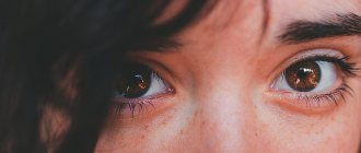
- Freckles are small rashes that are considered by some as a cute feature, by others as a cosmetic defect. During the cold season, freckles may disappear.
Freckles on the face
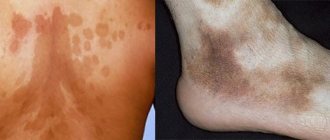
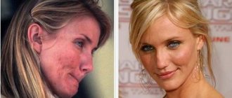
- Chloasma is hyperpigmentation of the skin in the form of large spots of light brown, brown or brown color with a smooth surface. Most often, chloasma is located on the face, but it can also occur on other parts of the body.
Chloasma
- Nevi (birthmarks) are pigmented areas of skin with smooth edges. The color of moles varies from light to dark brown. If moles change shape and increase in size, this may be a sign of its degeneration into a malignant form.
Mole on the body
Where can brown spots be located?
Pigmented areas can be located throughout the body. The nature of the disease depends on the location of the defect.
Localization of spots is as follows:
- Face and area around the nose, chin and mouth. It manifests itself mainly in women experiencing reproductive disorders.
- Knee joints, toes. An indicator of thyroid imbalance. More often seen in men.
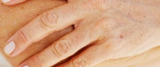
- Area of the palms, arms and wrists. Caused by senile changes in which metabolism is disrupted. The marks often have a bluish tint.
- Neck and shoulder area. At a young age, the pigment appears as a consequence of tanning; at an older age, after the age of 45, it indicates lentigo.
Diagnostics
A dermatologist provides consultations on skin diseases. When examining pigmentation, he will pay attention to details such as:
- size of education;
- surface of the stain (smooth, rough, etc.);
- color intensity;
- number of rashes and location;
- accompanying symptoms (itching, peeling, etc.).
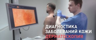
This will help the doctor make a preliminary conclusion about what it might be. For a more accurate diagnosis, you will need to take blood tests - general and hormone tests, and possibly do an ultrasound of the liver and thyroid gland. To examine the nature of brown spots on the body, a dermatoscope will be used and scrapings will be taken from the surface of the affected areas. If necessary, the dermatologist will prescribe the patient a consultation with an endocrinologist, gastroenterologist, gynecologist and other specialists.
Brown spots in a child
The skin of children has a thin stratum corneum, and the tissues are intensely saturated with blood vessels. All this, combined with a weak immune system, can lead to the formation of pigmented areas on the body. Hyperpigmentation often manifests itself as freckles, nevi (giant, flaming, melanocytic, non-cellular), in the form of hemangioma, congenital coffee spots.
Brown spots on the skin are caused by a hereditary form of the disease.
These types include:
- Neurofibromatosis type 1. Initially, café-au-lait spots are small in size, on the surface of which a cluster of freckles forms. The disease often occurs at an early age. The neglected process leads to oncological complications, on the basis of which diseases develop: respiratory tract cysts, growth retardation, formation of voids in the spinal cord, narrowing of the renal vessels, enlarged mammary glands.
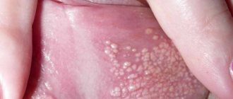
- Peutz-Jeghers disease. Pigmentation of this type affects large areas of the body, mucous membranes and leads to polyposis of internal organs. The formations are localized around the eyes, around the lips, in the anus and palms.
- Poikiloderma. Hereditary spots are observed in infants from 3 to 5 months. Initially, hyperemia occurs in the limbs, face, and neck. Then atrophic pigment changes form on the affected area.
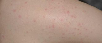
- Urticaria pigmentosa. This form of pigmentation can trigger the development of mastocytosis. A small dark red spot leads to the formation of vesicular inflammations filled with fluid. At the completion stage, the bubbles open, and in their place brown areas remain, which disappear after a few months. If left untreated, urticaria can cause disability or death. Pathology can be triggered by climatic conditions, stress, exposure to ultraviolet radiation, weak immunity, infectious and inflammatory processes.
Older children are susceptible to mastocytosis, allergic pigmentation, intradermal and borderline pigmentary nevus, as well as impaired melanin production (hypomelanosis). Deviations can manifest themselves in the form of diseases: albinism, vitiligo, Ito hypomelanosis.
A diagnostic examination is carried out using a dermatoscope, pigmentation is examined under ultraviolet radiation (Wood's lamp). In serious cases, computer diagnostics and biopsy tissue analysis are performed, followed by histological examination.
Treatment of pigment spots in children does not involve the use of lightening and whitening preparations containing hormonal components. At the age of 6-7 years, it is allowed to treat pigmented areas with parsley, lemon, and cucumber juice. If removal of the formation is required, then surgical method, laser therapy, electrocoagulation or cryotherapy are used.
How to treat brown spots
It is not always possible to cure pigmentation with medication, but modern means can make the spots less noticeable. You can purchase several types of whitening preparations at the pharmacy, some of them are presented in the table.
| Type | Name |
| Antifungal agents | Sulfur or zinc ointment, Clotrimazole |
| Antibacterial agents | Salicylic and syntomycin ointment |
| Exfoliates and reduces melanocyte production | Salicylic-zinc paste, folic, azelaic, kojenic acid. |
| Cream | Melanativ, Achromin |
| Cosmetical tools | Bodyaga forte, Boro plus cream, Clearwin, Biocon cream, Vitex mask. |
The desired result can be achieved by using the listed remedies for about 4-8 weeks.
Why do brown spots appear?
Brown spots on the skin are a result of increased production of melanin in the skin of the body. When the level of melanocytes increases, it leads to pigmentation disorders. This reaction is usually caused by an imbalance in metabolic processes and contributes to the development of skin disease.
An imbalance of pigments is associated not only with internal abnormalities in the functioning of organs, but is also provoked by stress and environmental factors.
The defect is caused by the following reasons:
- Intensive production of melanin under the influence of ultraviolet radiation, especially in fair-skinned people.
- Hormonal disorders, pregnancy, PMS, childbirth. Areas of skin may darken during pregnancy in the area of the nasolabial folds on the face. The deviation is caused by hormonal changes in the body and usually disappears after childbirth in a few months.
- Genetic factor (hereditary transmission).
- Age-related changes associated with deterioration of body systems, decreased tissue regeneration, and slower metabolism.
- Stress and depression disrupt the production of hormones and cause pigmentation.
- Chemical and thermal burns, wounds and blisters leave a brown mark on the body.
- Advanced form of acne, boils.
- Deficiency of microelements and vitamins (especially vitamin C, copper).
- Uncontrolled use of hormonal medications.
- Cosmetic procedures including acid peels, Botox, hyaluron, chemical cleansing, mesotherapy. Activities can injure the skin layer and stimulate increased melanin production.
- Diseases of the liver, stomach, kidneys, gallbladder, adrenal glands.
- Allergic reactions to cosmetics, certain foods.
Salon treatments for hyperpigmentation
Salon therapy sessions are recommended as the fastest way to eliminate pigment disorders.
Cosmetologists can offer several ways to remove stains:
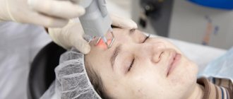
- Laser grinding. It is considered a highly effective procedure. The session is carried out mainly for the face. Under the action of a laser beam, melanin is destroyed, the problem area turns red, and then the skin becomes its normal color.
- Chemical peeling. It will help radically solve the problem of brown spots using an acidic composition (salicylic or triacetic). After 3-10 sessions, a significant result is noticeable.
- Phototherapy. Thanks to pulsed waves, the skin is lightened, the result is noticeable from the first use. Infrared radiation destroys melanin and does not damage the epithelium.
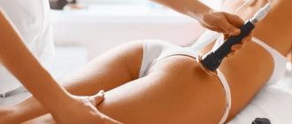
- Mesotherapy. The procedure consists of subcutaneous administration of a drug according to an individual prescription. In addition to the brightening effect, the technique rejuvenates and is considered less traumatic than laser resurfacing.
Despite the advantages of the procedures, there are contraindications that should be reviewed during consultation with a specialist.