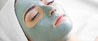
Features of the action of silicone gel against scars
Silicone gel has been used for many years to treat burns and heal post-surgical scars. The growing popularity of surgical plastic procedures and the development of cosmetology have led to the use of silicone not only for medical, but also for aesthetic purposes.
offer
For a wound to heal, it requires an environment that is neither too dry nor too wet. Silicone gel provides this environment, allowing the skin to breathe and preventing it from drying out. It stimulates fibroblast production while keeping collagen production under control and preventing scar tissue from growing. Silicone coating on the scar helps prevent bacterial infection, which can cause excessive collagen production and lead to the formation of keloid scars.
Wound healing requires not only the production but also the breakdown of collagen in the skin using natural chemicals called growth factors. Silicone can modulate the levels of various growth factors, controlling collagen production by maintaining the balance that is necessary for wound healing. Its use also helps reduce the discomfort and itching that often occurs during wound healing.
Biological effects of silicone gel
Since silicone coatings were first used to treat hypertrophic and keloid scars, they have proven to be effective and safe1,2.
Depending on the design of the study, applying a silicone coating to the scar area improved its condition in 60% - 100% of cases1–14 in terms of redness, itching, changes in the thickness and structure of scar tissue. As a rule, the first changes manifested themselves in tissue density, followed by changes in the color and height of the scars protruding above the surface of the skin7. Although silicone coatings are widely used in the treatment of hypertrophic and keloid scars, and much research has been carried out over the past two decades, the mechanism of action of these materials is still poorly understood1. Explaining this process seems all the more difficult since the pathogenesis of the formation of hypertrophic and keloid scars is not fully understood2. In addition, the use of a number of interchangeable terms makes it difficult to compare treatment efficacy results across studies. However, criteria for the differentiation of rough scars have recently been adopted5,16, and it has also been suggested that immunological mechanisms influence the process of their formation and development7–21. These observations demonstrated the presence of immune cell infiltrates in hypertrophic and keloid scars and allowed us to characterize their immunophenotypic properties, indicating a delayed type of immune response16,22. Hypertrophic and keloid scars exhibit different immunophenotypic profiles, and the amount of immune cell infiltrate is associated with age and clinical behavior of the scars22. There are reports that the use of silicone gels softens and smoothes the surface of excess scars in a shorter period of time than does the use of pressure dressings. The results obtained are not affected by compression, temperature or oxygen pressure4,5,8,23. However, there are also indications that temperature differences of less than 1°C observed with silicone coatings can have a significant effect on collagenase kinetics and significantly alter the scarring process1,24. Elasticity, measured by elastometry, is significantly increased when silicone gel sheets are used on hypertrophic scars compared to control sites8,12. In a recent study, we evaluated the effectiveness of silicone gel sheets in the treatment of hypertrophic and keloid scars, using objective measurements to confirm subjective assessments (biopsy with histological and immunohistochemical analysis of the samples). Quantification was performed by measuring tissue thickness using ultrasound and laser Doppler perfusion. Preliminary results indicated a significant reduction in the thickness of hypertrophic and keloid scars under the influence of silicone. The average % change in the total thickness of the scar also indicates its significant reduction compared to the initial level. There was also a decrease in scar perfusion, although these differences were not statistically significant, especially in the case of hypertrophic scars. Biopsies were performed in patients who consented to the procedure at baseline and 12 weeks after the start of therapy. There was a decrease in the number of spindle cells and an increased level of lymphocytes, which is apparently associated with the pronounced expression of CD11a/CD18 adhesion molecules (LFA-1). This finding suggests that silicone coating causes significant changes in the cellular infiltrate in hypertrophic and keloid scars25. Apparently, silicone does not penetrate into tissues after application of the gel to the skin, although this fact needs further clarification. Silicones are synthetic polymers whose supporting structure contains repeating silicon-oxygen bonds and organic groups directly bonded to the silicon atom. Elemental silicon is used as a feedstock in the production of many silicone-containing products. The most common synthetic polymer is polydimethylsiloxane (PDMS). Depending on the length of the polymer chain and the presence of cross-links, individual classes of silicone products are distinguished. Silicone gel sheets consist of a gel and a supporting elastomer membrane.
The gel has a small number of cross-links. In PDMS, cross-links are formed by vinyl and hydrogen groups bonded to individual silicon atoms in the presence of a catalyst. This polymer network is saturated with liquid PDMS polymer, forming an adhesive and viscous shapeless mass, the properties of which depend on the extent of the cross-links and the volume of added liquid. The elastomers consist of liquid PDMS polymer, have cross-links similar to those present in the gel, but much longer, and the content of free liquid polymer is very small. In addition, special forms of amorphous silicon compound account for approximately 30% of the total weight.
| FIGURE 1. A small number of CD68+ macrophages are present in the dermis 3 months after keloid excision (APAAP method, original magnification x100). | FIGURE 2. A large number of CD68+ macrophages are present in the dermis after removal of the keloid and application of silicone sheets for three months (APAAP method, original magnification x100). |
There have been no reported cases of inflammation or rejection reactions to the presence of a foreign body in scars that were covered with silicone gel, suggesting that silicone does not penetrate the treated tissue8. In addition, traces of the substance are not detected in the area of application of the silicone-containing dressing23. However, the release of silicon compounds from the surface of silicone wafers and their distribution into the skin has been demonstrated in in vitro experiments. In this case, silicon compounds, after distribution in tissues, may have some pharmacological effects on them. The mechanism of action of silicone coatings may be to create conditions of occlusion and hydration, which in turn promote changes in the condition of scar tissue. A number of studies have demonstrated that the use of silicone coatings causes hydration of the stratum corneum of the skin. In addition, during in vitro experiments it was noted that it was hydration, and not silicone, that suppressed the proliferation of fibroblasts and their production of collagen. The state of occlusion causes the appearance of an increased number of mononuclear cells in the epidermis. Some researchers have also reported the activation of the Langerhans cell system (Langenhars cells are able to reach the granular layer with their dendrites and come into contact with mononuclear cells) and their appearance in the dermis after occlusion2. Treatment of hypertrophic scars using occlusive dressings containing silicone and hydrogel caused changes in cytokine mRNA levels. Increased levels of interleukin-8 (IL-8), basic fibroblast growth factor, granulocyte-macrophage colony-stimulating factor, and decreased levels of transforming growth factor and fibronectin were observed after both types of dressings were used. Despite this, only hydrogel dressings showed significant differences in IL-8 and basic fibroblast growth factor levels relative to untreated scar areas. Significant changes in IL-8 and fibronectin levels were observed with hydrogel dressings, whereas significant changes were only seen in fibronectrin with silicone gel dressings compared to normal skin. It is likely that occlusion and hydration may have biological effects on hypertrophic and keloid scars with and without silicone. However, it should be noted that the effect of silicone is probably due not only to the effect of hydration, but is also associated with a number of other mechanisms. For example, there are references to the fact that static electricity created by the negatively charged components of the silicone coating contributes to the involution of hypertrophic and keloid scars, while causing a reduction in the duration of their therapy. The use of silicone in the form of gels/plates has proven effective not only in therapy, but also in the prevention of pathological scarring. Application of silicone coatings after surgical resection prevented the development of hypertrophic and keloid scars in 75% - 85% of cases1,2,9,23. A previous report presented data from a study conducted among patients who had relapsed after previous surgical removal of keloids. In patients of the first group, keloid scars were removed surgically without subsequent administration of a preventive course; patients in the second group used silicone for 3 months after removal of the scars. Scars treated with silicone coating had a significantly higher rate of complete remission. In addition, higher numbers of CD36+ dendritic cells and CD68+ macrophages were found in silicone-treated scars compared with their baseline condition and compared with scars that were not treated with silicone (Figures 1, 2). This fact indicates that the use of silicone coating in the form of gel or sheets may induce the process of scar tissue remodeling. In conclusion, I would like to note the need for new research to further study the mechanisms of action of silicone coating (gel), which, in fact, is the only non-invasive method that has proven its effectiveness and safety in the prevention and treatment of hypertrophic and keloid scars.
LIST OF SCIENTIFIC PUBLICATIONS
- Lyle WG. Silicone gel sheeting, Plast Rec Surg 2001; 107:272-5
- Niessen FB, Spauwen PHM, Schalkwijk J, Kon M. On the nature of hypertrophic scars and keloids a review. Plast Rec Surg 1999;104:1435-48
- Perkins K, Wallis KA. Silicone gel: new treatment for burn scars and contractures. Burns Incl Therm Inj 1983; 9: 201-4.
- Quinn KJ, Evans JH, Courtney JM, Gaylor JD, Reid WH. Non-pressure treatment of hypertrophic scars. Burn Incl Therm Inj 1985; 12: 102-8.
- Quinn KJ. Silicone gel in scar treatment. Burn Incl Therm Inj 1987; 13(Suppl):S33-40.
- Ohmori S. Effectiveness of silastic sheet coverage in the treatment of scar keloid (hypertrophic scar). Aesth Plast Surg 1988; 12:95-9.
- Mercer NS. Silicone gel in the treatment of keloid scars. Br J Plast Surg 1989 42 83-7.
- Ahn ST, Monafo WW, Mustoe TA. Topical silicone gel: a new treatment for hypertrophic scars. Surgery 1989; 106: 781-7.
- Ahn ST, Monafo WW, Mustoe TA. Topical silicone gel for prevention and treatment of hypertrophic scars. Arch Surg 1991; 126:499-504.
- Gold MH. Topical silicone gel sheeting in the treatment of hypertrophic scars and keloids. A dermatologic experience. J Dermatol Surg Oncol 1993; 19: 912-6.
- Gold MH. A controlled clinical trial of topical silicone gel sheeting in the treatment of hypertrophic scars and keloids. J Am Acad Dermatol 1994; 30: 506-7.
- Carney SA, Cason CG, Gowar JP. Cica-Care gel sheeting in the management of hypertrophic scarring. Burns 1994; 20: 163-7.
- Dockery GL, Nilson RZ. Treatment of hypertrophic and keloid scars with silastic gel sheeting. J Foot Ankle Surg 1994; 33: 110-9.
- Gibbons M, Zuker R, Brown M, Candlish S, Snider L, Zimmer P. Experience with Silastic gel in pediatric scarring. J Burn Care Reabil 1994;15:69-73.
- Ehrilich PH, Desmouliere A, Diegelmann RF, Cohen IK, Compton CC, Garner WL, Kapanci Y, Gabbiani G. Morphological and immunochemical differences between keloid and hypertrophic scar. Am J Pathol 1994; 145: 105-13.
- Santucci M, Borgognoni L, Reali UM, Gabbiani G., Keloids and hypertrophy scars of Caucasians show distinctive morphologic and immunophenotypic profiles. Virch Arch 2001; 438:457-63.
- Placik O, Lewis VL, Immunological associations of keloids. Surgery 1992; 175:185-93.
- Shetlar MR, Shetlar CL, Hedricks L, Kischer CW. The use of athymic nude mice for the study of human keloids. Proc Soc Exp Biol Med 1985; 179:549-52.
- Kischer CW, Pindur J, Shetlar MR, Shetlar CL. Implants of hypertronic scars and keloids in to the nude (athymic) mouse: viability and morphology. J Trauma 1989; 29:672-7.
- De Limpens J, Cormane RH. Keloids and hypertronic scars: immunological aspects. Aesth Plast Surg 1982; 6:149-52.
- Castagnoli C, Stella M, Magliacani G, Alasia ST, Richiardi P. Anomalous expression of HLA class 2 molecules on keratinocytes and fibroblasts in hypertrophic scars consequent to thermal injury. Clin Exp Immunol 1990; 82:350-4.
- Borgognoni L, Pimpinelli N, Mrtini L, Brandani P, Reali UM. Immuno-histologic features of normal and pathologic scars: possible clues to the pathogenesis. Eur J Dermatol 1995; 5:407-12.
- Fulton JE, Silicone gel sheeting for the prevention and management of evolving hypertrophic and keloid scars. Dermatol Surg 1995.
- Kreiger LM, Pan F, Doong H, Lee RC. Thermal response of the epidermis to surface gels. Surg Forum 1993; 44:738-42.
- Borgognoni L, Mrtini L, Brandani P, Magini B, Reali UM. Objective measurements used in the investigation of the effects of silicone gel sheeting in the treatment of HS and K. Wound Rep Reg 2000; 8:
Indications for use of silicone gel
Silicone gel is effective for treating different types of scars:
KK Adapt. 5 paragraph
- burns;
- keloids;
- hypertrophic;
- atrophic;
- post-acne;
- common surgical scars;
- scars from cosmetic surgical procedures.
Silicone gel helps in the epidermal repair process by slightly increasing the temperature of the skin and creating continuous light pressure on the wound.
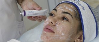
Scar preparations
Most brands of silicone gels contain long chains of silicone polymer (polysiloxanes) and silica, usually mixed with a volatile compound that evaporates, drying out the surface of the skin within minutes of application. They also contain surfactants. These are essential oils, vitamins and minerals that help moisturize, nourish and soften the surface of the skin. They increase blood circulation, accelerate local metabolism, promoting the removal of tissue decay products.
After drying, the gels create a protective transparent barrier that provides a therapeutic effect. They are easy to use and safe even for children's sensitive skin.
Silicone gel for scars Contractubex
This is one of the most famous scar medications. The gel contains allatoin, sodium heparin, onion extract. It removes dead skin particles and stimulates the production of new cells. Improves hydration, softens scar tissue and normalizes the formation of fibroblasts.
Contractubex gel is started to be used after the wound has healed. The damaged area is treated 3 times a day. Fresh scars smooth out within a month. To improve the condition of an old scar, you must use the gel for at least 6 months. Longer use should be agreed with your doctor.
offer
Silicone gel for scars Kelo-kot
Kelo-Kot comes in both gel and spray form and is considered the most effective scar treatment available today. Its basis is silicone dioxide and polysiloxane (a combination of silicone and oxygen). This gel is very gentle on the skin, so it can be used to remove scars on the face and sensitive skin. The drug should be applied once a day and not washed off. After it dries, a transparent film forms on the skin, which has a therapeutic effect throughout the day.
Dermatix gel
Dermatix is a preparation that contains inert surface-acting silicone. The gel accelerates the healing process of postoperative scars and reduces the severity of old scars. Apply the product 2 times a day to cleansed skin.
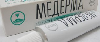
Silicone gel for scars Mederma
The effectiveness of the gel is ensured by its rich composition:
- xanthan;
- cepalin;
- allatoin;
- sorbic acid.
This drug not only helps to resolve scars. It is used to combat stretch marks and marks after tattoo removal. The gel is recommended for use after plastic surgery. It should be applied 4 times a day. The treatment period is 3–6 months, depending on the type of scar tissue.
Fermenkol
This gel is recommended for the correction of post-burn scars, scars from mechanical injuries, atrophic post-acne scars. Fermenkol effectively softens the skin, relieves itching and pain. You can begin gel treatment after 3–4 weeks of wound healing.
Dermatix gel 15g
Silicone gel DERMATIX to prevent scarring
DERMATIX – silicone gel for preventing the formation of scars immediately after wound healing and correction of immature protruding scars.
What is Dermatix gel?
DERMATIX is a transparent, quick-drying gel that helps maintain uniform skin moisture and has a corrective effect on scars resulting from injuries, burns, surgical interventions and other skin damage.
DERMATIX has the ability to smooth and soften raised scars, relieve associated pain, itching and discomfort, and reduce skin redness.
DERMATIX is easily applied to all areas of the skin, including the face, joints and folds, dries quickly and forms an invisible protective “film”.
What is Dermatix gel used for?
DERMATIX gel is used to treat and prevent the formation of hypertrophic and keloid scars (after surgery, burns or other injuries) in the early stages immediately after wound healing.
The gel can also be used for raised immature red scars that are in the process of formation and up to 2 years after wound closure.
How is Dermatix gel used?
DERMATIX gel should be applied 2 times a day (morning and evening), following the instructions below:
• Rinse the treated area of skin with water and pat dry. DERMATIX should only be applied to clean and dry skin.
• Using gentle massage movements, rub a small amount of gel into the scar. If you apply too much gel, the excess should be removed with a napkin so as not to stain your clothes.
• After Dermatix gel has completely dried, you can apply cosmetics.
The minimum duration of the preventive course is at least 2 months; in case of complicated scarring, longer use may be required.
DERMATIX can be used in pediatrics.
In what cases should Dermatix gel not be used?
• DERMATIX gel should not be used on open, unhealed wounds.
• Avoid contact of the gel with the mucous membrane and areas of the skin located in close proximity to the eyes.
• DERMATIX should not be applied over any dermatological preparations without consulting a physician.
Are there any possible side effects when using DERMATIX gel?
In rare cases, the use of the gel may cause redness, pain or irritation of the skin. If this worries you, or you notice that other undesirable manifestations are developing, consult your doctor.