This is a gland that secretes milk, a characteristic feature of animals belonging to the class of mammals. The mammary glands (mamma; synonymous with the mammary gland) are a paired glandular organ that produces milk in women after childbirth; in men it remains underdeveloped and does not function. Its secret is breast milk, the natural food of young mammals in the initial postpartum period of development. Breast diseases are currently “younger,” which makes the problem of early and modern diagnosis even more relevant in the practice of mammologists and gynecologists.
Outside the lactation period, it has an average diameter of 10-12 cm and a thickness of 2-3 cm. The weight of the gland in girls ranges from 150-200 g, during the lactation period 300-900 g. In most young healthy women, it is elastic and has hemisphere shape.
Evolutionary purpose of the mammary glands
The mammary gland in women is an important organ of the reproductive system, which begins to function at a certain period of life. Its main task is to produce milk to feed the baby after birth. This feature places a person in the class of mammals - according to the scenario planned by nature, the cub is fed milk for several months, receiving all the most useful things from the mother.
The gland can fully function only after the birth of the child; its cells “mature” and begin to produce milk. This feature depends on the hormonal background, the variability of which affects the qualitative composition of its tissue.
Interesting: the structure of the mammary gland does not differ between boys and girls before puberty. When puberty occurs, under the influence of female hormones, its tissue begins to grow and develop. In men, the gland remains in an atrophied state for life.
Many people believe that men do not get breast cancer. But that's not true.
For many years now, most of our patients undergo an annual routine examination - clinical examination. We recommend that women and men begin medical examination as early as possible and carry it out regularly.
For many years now, most of our patients undergo an annual routine examination - clinical examination. We recommend that women and men begin medical examination as early as possible and carry it out regularly.
The examination includes a block of tests, ultrasound examinations, ECG and consultations with specialists. The examination is aimed at identifying both risk factors and early signs of the development of diseases: heart, liver, blood, genitourinary organs, oncological pathology, and so on.
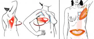
The statistics are disappointing. Cancer and cardiovascular diseases are becoming younger and remain firmly in first place in terms of morbidity and mortality. And you should know that many conditions can be prevented or treated in the early stages. And that is why the issue of early diagnosis is so acute. There are three principles here: regular self-examination (see pages on our websites: breast self-examination , testicular self-examination ) + annual examination + contacting a doctor when the first symptoms of disease are detected.
I suggest you read the message on the topic of breast cancer in men.
Many people believe that men do not develop breast cancer. But that's not true.
Although this type of cancer is extremely common in women, breast cancer in men is extremely rare.
The breasts of an adult man are similar in structure to the breasts of girls before puberty, and consist of several ducts surrounded by fatty and other tissues. In girls, this tissue grows and develops during puberty under the influence of hormones, but in men it does not.
Risk factors for developing breast cancer in men
- Age from 50 to 70 years
- According to the literature, the risk is higher in African-Americans than in Europeans.
- Gynecomastia and obesity, as well as the conditions under which they develop (Klinefelter syndrome, severe liver disease, taking certain medications)
- Heredity
- Radiation exposure
- Treatment with estrogens
- BRCA2 gene mutations
Thus, according to various authors, some patients associate the disease with trauma, some patients have relatives with this disease, almost half of the patients suffered from gynecomastia.
How serious is this disease?
In the early stages, the disease is treatable. The problem is that breast cancer is diagnosed later in men than in women. This is due both to the fact that this type of cancer is very rare, and to the fact that men are less likely to pay special attention to this area of the body, going to the doctor with more severe symptoms (bleeding, changes in the skin of the breast).
Symptoms of breast cancer in men
- Swelling
- Palpable mass inside the chest
- Wrinkling of breast skin
- Nipple changes (redness, shrinkage)
- Pain in the breast and nipple area
- Nipple discharge
Diagnostics
First of all, at a medical appointment, examination and palpation of the breast, lymph nodes, and assessment of the condition of the liver are carried out. The doctor asks in detail about heredity and identifies the presence of risk factors. If the doctor detects any changes as a result of the examination, a full examination is prescribed.
Mammography is an X-ray examination of the breast.
Ultrasound examination
Biopsy is the removal of a sample of breast tissue for histological examination (examination under a microscope).
Tests - determination of tumor markers, estrogen/progesterone receptors, genetic testing. The main markers are CEA and SA-15-3.
Treatment
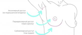
Surgical – in men, a radical mastectomy is most often performed, that is, removal of the entire mammary gland, part of the chest muscles, and axillary lymph nodes. The removed tissue is sent to the laboratory for histological examination.
Radiation therapy – to stop the growth and division of cancer cells.
Chemotherapy is carried out using anticancer drugs.
Hormonal therapy is carried out with antiestrogens and antiandrogens if the tumor cells have receptors for estrogen/progesterone.
Panel of tumor markers for men for men Spermatology CA-15-3, breast diseases in men CA-15-3
Breast cancer Tumor markers Use of some markers in the diagnosis of malignant neoplasms
Tags:
tumor markers, breast cancer, medical examination, cancer
Back to section
Features of anatomy
In an adult woman, the mammary gland has a structured structure, consisting of several types of tissue, each of which performs its own function. Let's take a closer look at the anatomical features:
- The outside of the gland is covered with skin, on which the juice area is located. It is rich in pigment cells, which plays an important role in feeding the baby - the baby instinctively finds the nipple, since its surface is slightly different from smooth skin. The areola also contains a large number of nerve endings.
- The parenchyma of the breast has a segmental structure - dense strands extend from the skin, which form large cells (15-20), surrounded by fatty tissue. They contain lobules of the mammary gland, separated by connective tissue.
- Each lobule consists of glands, inside of which there are many alveoli that produce milk. Convoluted tubules extend from them, they gradually unite and form the nipple opening.
- The entire anatomical structure is supported by a special cord called the ligament that supports the mammary gland. Underneath it there is a layer of fat that prevents soft tissue injuries.
Normally, there should be no lumps in the breast; the entire structure is soft and can be painlessly palpated. The mobile ligament provides slight displacement without noticeable discomfort. Proportional sizes mainly depend on the volume of adipose tissue that fills the free space between the cells.
Women's breasts. Breast structure
The female breast is a complex organ, built to provide optimal conditions for performing its main physiological functions: milk production and feeding the baby. The breast consists of skin, under which the gland itself is hidden, as it is also called, glandular tissue - the very organ in which milk is formed. Glandular tissue (gland) is attached by connective tissue to the muscles of the chest. Around the glandular tissue, between its lobes lies fat - adipose tissue.
The amount of fat in a woman's breasts varies widely. Some women's breasts are composed almost exclusively of fat. As a result, their breasts can vary greatly in size as their body weight fluctuates.
Some women have much more glandular tissue than fat, and the size of their breasts is practically independent of diet and weight. If the growth of adipose tissue can be accelerated by abundant nutrition, then the growth of glandular tissue is partially controlled by hormones. This explains why breast size can change during the menstrual cycle or after menopause.
Beneath the mammary gland lies the pectoralis major muscle. The chest is, as it were, attached to this muscle, but in the chest itself, contrary to popular belief, there are no muscles, so it is impossible to increase breast size through exercise. You can tighten the surrounding muscles, but this will only lead to an increase in the volume of the torso and will not affect the size of the breast itself. Of course, it is impossible to tighten sagging breasts through exercise.
There is a common belief that after plastic surgery, the breasts, as a rule, lose reflex responsiveness. Allegedly, during the incision, the nerves are cut, as a result of which the breast loses sensitivity and ceases to be an erogenous (especially sensitive zone). This is not entirely true. The 4th intercostal nerve is responsible for the sensitivity of the nipple and areola. It passes at the level of the axillary line, branches into two parts and, passing along the circumference of the chest, enters the gland tissue.
This nerve is not cut during surgery, however, if the implant is not placed correctly, or if the implant is too large, this nerve can become trapped between the prosthesis and the chest and become pinched. If the 4th intercostal nerve becomes pinched or damaged, the breast may lose sensation. This complication occurs in 21% of women. However, first-class specialists should not have such defects.
It would be wrong if scientists remained aloof from such an important issue as the correct volume and shape of the female breast. The average parameters of the “ideal” breast were calculated through long measurements of classical and modern sculptures and live models. The basis was a woman 162 cm tall, aged 17–18 years. After much debate, most authors accepted the following dimensions: the diameter of the areola averages 3.7 cm (37 mm) and varies from 3.5 to 4.5 cm. The distance between the collarbone and the nipple averages 21 cm. The distance from the inframammary fold to the nipple is approximately 7 cm.
However, when analyzing the real state of affairs, one can see how far we are from ideals and how contradictory the tastes of various authors regarding the size and volume of the mammary glands are. Some researchers suggest that the volume of a woman's breasts be considered optimal as 275 cubic centimeters (size C), and larger volumes are considered hypertrophy, i.e. increase relative to the average norm. However, in the search for the ideal, it is necessary to take into account such body features as height, weight, and figure features. For example, fragile women with a height of 152 - 177 cm and a weight of 45 - 54 kg look harmoniously developed, having a chest volume of 150 - 200 cubic meters. cm (size B).
In fact, the ideal shape and size of a woman's breasts are determined in accordance with racial and, in many ways, individual, social and aesthetic ideas. And, sometimes, it is completely incomprehensible why some women deliberately choose disproportion and achieve success.
There are contraindications. Read the instructions or consult a specialist.
The structure of the mammary gland during puberty
In girls, the mammary gland is in a embryonic state. There is skin on the outside and a small layer of fat on the inside.
Characteristic age-related features appear during the production of female sex hormones:
- milky tubes appear in the lobules of the mammary gland, which begin to grow and branch;
- when the ducts finish growing, alveoli form at their ends;
- cells of adipose and connective tissue begin to actively divide, as a result of which the mammary gland increases in size.
The result of all these processes is breast growth. The body prepares for pregnancy, and subsequently the glandular and adipose tissue undergoes certain cyclic changes.
Embryological and comparative anatomical data.
The first rudiment of the milk secretion organs appears in the form of a paired epithelial strip on the ventral wall of the body. This rudiment is called the milky stripes. The milky stripes thicken and turn into milky plates (milky lines). The individual projections developing from them are called milk tubercles (primary nipples). These latter, becoming flatter and growing deeper like protrusions, give this name. milky points, or points; through the immersion of individual glandular areas, they lead to the formation of milky pockets.
The first germ of mamma
Guldberg (1899) found in dolphin embryos, 18 millimeters long, in the form of vaguely demarcated elevations of the epidermis, which in this place forms a crescent-shaped protrusion. The rudiment appears just at the time when the temporary hind limbs begin to show a tendency to disappear.
In cattle embryos, according to G. Burckhardt, “accessory” or “anal” nipples are found very often in both sexes (in approximately 37% of cases). They are always located between the normal nipples (on one or both sides), or behind the back pair of normal ones; but they are never ahead of the first pair. The mammary organs of modern cattle are subject to the process of caudocranial reduction.
Both in humans and in cattle, they distinguish between hypermastia, with all the signs of a normal, but only very small, mammary organ, and hyperthelia (false nipples).
False nipples and mikromammae
disappear, for the most part, already before birth.
In his article on the morphology and history of the development of normal and accessory mammary glands, G. Schickele
) (1889) comes to the following conclusions: in some mammals (mouse, rat, rabbit, cat) the nipples are arranged in 2 groups, which are characterized by cranial and caudal convergence, as well as the presence of a typical significant gap between the most caudal and the most cranial pair of glands.
Accessory areas of glands in these animals, of course, occur only to a limited extent. But broad-nosed monkeys have extra nipples more often than narrow-nosed monkeys. At Zebus
this condition seems to be observed very often.
In guinea pigs and mice, the milk line is the starting point for the formation of glands. Adult guinea pigs have never had more than 2 teats. But embryonic accessory rudiments of the mammary glands are found in varying numbers (from 2 to 10); but they are at a lower stage of development than the rudiments of the main glands ( Ztschr. fur Morph. u. Antroph. 1899
).
Schmidt, H, Uber normale Hyperthelie menschlicher Embryonen. Anat. Anz. XI, 1896; and Morphologische Arbeiten, herausg. v. G. Schwalbe. 7 Bd.
1896.
Humans do not have a milk line as noticeable from the outside as in mammalian embryos, or it is present only for a short distance. But microscopic examination reveals growths of the epithelium and the rudiments of the mammary glands. In one case there were 8 additional rudiments on each side, 4 above and 4 below the main rudiment. In other cases, a rudiment of 7 to 14 parts was found on one side. The cranial primordia were mostly oriented laterally, while the caudal primordia were oriented medially with respect to the normal primordium. In the lower abdomen, the rudiments were completely absent.
Callius ( Anat. Hefte, Bd. III
, 1897) found in a 15 mm human embryo a milky line 1.5 mm long.
Regarding the comparative anatomy of the mammary glands, the reader can find detailed data in the relevant textbooks. We will only point out here that Monotremata has a paired and “glandular area,” which generally corresponds to areola mammae
higher, is the only external apparatus; The glands here are built according to the tubular type. In echidna, such a device is hidden in a skin pocket. Alveolar mammary glands first appear in marsupials (Gegenbauer).
Bonnett, Die Mammaorgane. Ergebnisse der Anat. von Merkel und Bonnett. Bd. II and VII. — Breslau , E., Beitrage zur Entwicklungsgeschichte der Mammarorgane bei den Beuteltieren. Zeitschr. f. Morph. u. Antrop. IV
, 1902.
The pouch arises through the fusion of small pockets (marsupial pockets), each of which contains one lacteal rudiment (mammary pocket). Marsupialia bag
is homologous to the echidna bursa, in the same way the marsupial pocket of the first is homologous to the corresponding formation in the second;
the mamillary pouch of Marsupialia
corresponds to the glandular field of the echidna.
Marsupial pouches in marsupials can be demonstrated even in placental animals. They correspond to the pockets that surround the Murinae
nipples. In all mammals, the mammary glands arise in a homogeneous manner and should be classified as tubular skin glands. The possibility of diphyletic origin must be excluded.
Unger (1898) also subscribes to this conclusion. The mammary glands are derived from the glomerular glands. Normal, comparative and pathological anatomy, phylogeny and ontogeny unanimously point to precisely this origin of the mammary glands. Neither their structure nor their function gives us the right to compare them with the sebaceous glands.
H. Eggeling
) in his work on the skin glands of Monotremata (Verh. anat. Ges. 1900) criticizes previous attempts to subdivide the skin glands.
As a principle of division, one must use, firstly, the ratio of the epithelium to the lumen, and, then, the method of secretion formation. All glomerular glands, and together with them the mammary glands of higher mammals, should be considered as constantly canalized skin glands secreting in the intravital state; the sebaceous glands and peculiar glandular organs of reptiles should be considered temporarily canalized, secreting through necrobiosis by the skin glands; their secretion is released necrobiotically, as the secreting cells die. v.
Runge, G., Die zunehmende Unfahigkeit der Frauen, ihre Kinder zu stillen. Munchen., E. Reinhardt , 1900.
The effect of menstruation on breast anatomy
In an adult woman, breast tissue is constantly exposed to hormones, their concentration varies depending on the phase of the cycle. Clinically, two periods can be distinguished during which changes occur at the cellular level:
- Temporary hypertrophy - in the second phase of the cycle, progesterone is produced in high concentrations. Under its influence, milk tubes begin to grow and alveoli appear. By the end of the month, the described changes reach their peak, the breasts may increase slightly in size, and sometimes pain occurs.
- Reduction - if fertilization does not occur, against the background of a decrease in progesterone concentration, the alveoli undergo reverse development. The body takes a short break to prepare for the next phase of the cycle.
Women's milk, lac feminum.
Due to the presence of numerous fat globules, it appears as a pure white or bluish-white liquid, is odorless, has a weak sweetish taste and exhibits a neutral reaction.
Its specific gravity is from 1028 to 1034, and its temperature at the time of separation is 38.C. The milk secreted in the first days after birth is transitional milk and is called colostrum puerperarum
;
it is, for the most part, thicker, yellower, grayer, sometimes even thinner than later milk. The fluid released from the breasts during pregnancy is called colostrum of pregnant women, colostrum gravidarum
, and over time gradually takes on the properties of
colostrum puerperarum
. Both of these fluids represent unripe milk and are characterized by the presence of a large number of large nucleated cells loaded with fat globules; they are called Donne bodies, or colostrum bodies. In all likelihood, these are nothing more than lymphatic cells loaded with fat. “Similar relationships are observed in mammals.
Formal components of mature milk. are numerous rounded fat drops, ranging in size from 2 to 5 microns, called milk globules, corpuscula lactis
, and a few lymphatic bodies.
Apparently, these fat globules are surrounded by thin protein membranes. When the milk sits, some of the fat globules separate from the liquid, rise to the top and form cream, cremor lactis
.
Milk completely freed from fat globules represents milk plasma; it also contains casein in solution. Due to the action of enzymes and acidification, milk curdles, i.e. casein falls out. The liquid remaining after casein is removed is whey, serum lactis
.
The most important chemical components of milk can be expressed in the following average figures.
Water 87.79%, solids 12.21%; casein and albumin 2.11% fat 3.79%; milk sugar 5.71%; salts 0.24%. Its other organic components are peptone, urea, and lecithin. Of the inorganic salts, human milk, according to Bunge, contains: potassium 0.0703%, sodium 0.0257%; lime 0.0343%; magnesia 0.0005%; iron oxide 0.0006%; phosphoric acid 0.0468%; chlorine 0.0445%; together this amounts to 0.2287%, while cow's milk contains only 0.8404%.
According to Bouchut, a cubic millimeter of milk contains, on average, 1,026,000 large and small milk pellets.
At the end of the lactation period or if feeding is not carried out, the gland undergoes reverse development, whereby the alveoli become smaller again, and their cavities, as well as passages and epithelial cells, are filled with fat drops and granular decay. During menopausal involution, the mammary glands gradually obliterate, and this process can even extend to the excretory ducts.
It is interesting that even the mammary gland of newborns is already able to produce secretions; that's the name. witches milk, lac neonatorum
. According to some researchers, this secretion is not real milk; but, in any case, both chemical and microscopic examination indicate a significant similarity between these two secretions. Barfour is also inclined to consider this secretion to be milk.
| Fig. 62. Network of lymphatic vessels on the anterior surface of the mamma; subareolar plexus; trunks . | |
Arteries originate from aa. intercostales
(
rr. mammarii mediales et latt.
), and also from
rr.
mammarii , belonging to
a.
mammaria interna [According to PNA -
a.
thoracica interna - IIH].
Arterial anastomoses, into which rr ultimately enter, are considered (without reason)
as an incidental reason for the stronger development of mamma .
mammarii , belonging to
mammaria interna
, with
aa.
uterinae (through aa. epigastrica superior and inferior).
Namely, a.
epigastrica inferior gives
a.
spermatica externa [According to PNA -
a.
cremasterica in men
and a.
lig. teretis uteri in women - IIH], and the latter anastomoses with a. uterina, as it goes to the uterus along lig. teres uteri.
The saphenous veins form the papilla mammae
polygonal network of anastomoses,
plexus venosus mamillae
(see section III.).
The saphenous veins stretch to neighboring large veins, even to v. cephalica
, while deep veins follow the arteries. Lymphatic vessels form a narrow-loop plexus in the skin covering the glands, especially in the areola. The interalveolar connective tissue also contains lymphatic vessels. Fig. 62.
Nerves are quite numerous in the outer skin, in the areola
and
papilla mammae
, but there are few of them inside the gland, and here they are mainly vascular nerves.
They start from nn.
supraclaviculares , from
rr.
cutanei antt. , belonging to
nn.
intercostales II-V-VI , pass through the skin radially to the nipple and in the form of
rami glandulares
, originating from
nn.
intercostales IV-VI , penetrate into the gland itself. Together with the arteries, the glands and their sympathetic plexuses penetrate inside.
In the papillae papilla mammae
tactile corpuscles are found; At the base of the nipple there are separate Vater-Pacinievsky bodies. Near the larger milky passages, V. Krause found end flasks.
The nipple is capable of erection and, when its cutaneous nerves are irritated, lengthens; but erection is determined only by the action of the corresponding smooth muscles, without any participation of the venous spaces.
Changes in the breasts during pregnancy
The mammary gland can function fully only during pregnancy and after childbirth. Natural physiological mechanisms are launched that ensure milk secretion.
The structural features are determined by three hormones:
- Progesterone - under its influence, the alveoli reach their final development and are ready for secretion. The milk ducts grow in the lobules, the glands prepare to produce milk.
- Prolactin - produced immediately after childbirth, triggers secretion in the alveoli and the release of colostrum through the ducts. It contains a large amount of vitamins, minerals and nutrients that a child needs.
- Oxytocin - supports the secretion and production of mother's milk for several months. Under its influence, the alveoli remain in a hypertrophied state until the end of feeding. When the hormone titer decreases, all structures of the mammary gland undergo reduction.
Against the background of all these changes, the mammary glands are exposed to a dozen hormones. The whole system works like a clock; even a small malfunction can lead to disruption of cell division in the breast and the appearance of problems.
Types of mammary gland structure
In women, the structure of the breast changes at different periods of life, sometimes the anatomy is influenced by the individual characteristics of the body. There are many classifications, we will note the gradation by type of fabric:
- Glandular type - occurs in young women and is considered a normal variant. Adipose tissue is present, but its proportion is small.
- The fat type of structure is typical for overweight women. It also occurs with a normal physique, when after 35-40 years the processes of reverse development begin in the chest and the glandular tissue is replaced by adipose tissue.
- Mixed type - intermediate. Characterized by an equal ratio of adipose and glandular tissue.
Pathologies of the mammary glands
If hormonal imbalances or abnormalities in physiology occur in the body, local diseases may develop. We list the pathologies in descending order of frequency of occurrence:
- mastopathy – the appearance of lumps in the breast;
- cancer - usually occurs as a complication from a previous disease;
- mastitis – inflammation in the breast tissue due to congestion;
- infections - often bacterial or fungal in nature.
It is very important to prevent the listed pathologies - these are regular examinations and palpation of the breast. Timely delivery and breastfeeding play an important role. You should also choose a comfortable bra made from natural materials.