For the treatment of varicose veins of the lower extremities, all conditions have been created at the Yusupov Hospital:
- Chambers with European level of comfort;
- Modern equipment from leading global manufacturers;
- Carrying out conservative therapy with the latest, most effective drugs;
- The use of traditional and innovative surgical techniques by phlebologists;
- Attentive attitude of medical staff towards patients and their relatives.
Hematomas and swelling of the leg after venous surgery can be a consequence of the operation or its complication. In the first case, the hematoma resolves on its own, and in the second, patients are prescribed treatment with modern drugs. If your leg swells after vein surgery, you should consult your doctor.
Causes
Hemorrhage usually occurs after injury. This could be a bruise of the skin, internal organs, a concussion or bruise of the brain, an injection with thin (sharp) objects. Sometimes blood leaves the vessels and pours into the skin and internal organs as a result of infections, autoimmune diseases, and poisoning. The occurrence of hemorrhages and bruises is promoted by increased fragility of blood vessels, fasting, lack of vitamins in food, high blood pressure, and congenital bleeding disorders.
At CELT you can get a consultation with a traumatologist-orthopedic specialist.
- Initial consultation – 3,000
- Repeated consultation – 2,000
Make an appointment
Symptoms
A bruise on the skin is always clearly visible. At first it has a purplish-blue color, and then begins to “bloom”, acquiring yellow and green colors. If a sufficiently large amount of blood accumulates under the skin, a protruding lump forms. At first it is very painful to feel, but later the pain goes away.
The outpouring of blood into the internal organs and into the substance of the brain is preceded by trauma. The main symptom is pain. In this case, the hematoma is not visible externally. If bleeding continues, the victim becomes pale, weak, and dizzy. With chronic internal bleeding, anemia comes to the fore. Bleeding in the brain is especially dangerous. Compression of brain structures may occur, sometimes leading to death.
4.Treatment
As shown above, the options for the clinical development and outcome of renal hematoma are so diverse that it is not possible to outline even the contours of a universal therapeutic approach. The diagnostic results and dynamic factors (reduction/increase in hematuria, improvement/aggravation of the general condition, presence/absence of signs of secondary infection, increase/decrease in blood pressure, etc.) determine the choice of protocol in each specific case with all its anamnestic and clinical nuances. In relatively mild cases, the situation can be resolved successfully with gentle or bed rest (for 7-10 days) with the exception of physical activity and constant monitoring of the condition, control of urination, pressure, body temperature and other indicators. To prevent infectious and inflammatory processes, antibiotics are prescribed; Hemostatic agents and local hypothermia (cold applied to the injured area) may be indicated. In case of a less favorable development of events, they act according to one or another protocol of surgical intervention (sometimes on an urgent or emergency basis), the options and methodological modifications of which are also numerous - from various organ-preserving operations to kidney removal.
Renal hematomas, in general, are dangerous not only due to their pronounced polymorphic clinical picture and unpredictable dynamics, but also due to long-term complications: the formation of stones, chronic nephrogenic arterial hypertension, pyelonephritis, etc. Therefore, even with a seemingly minor injury to the kidney - especially if it is accompanied by noticeable swelling, edema, persistent sharp pain, cloudy or red urine, high fever, general malaise - you should see a nephrologist, urologist or therapist as quickly as possible. The time factor in prognostic terms may be critical.
Predisposing factors
The formation of hematomas occurs after injuries, including pinching, blows, squeezing, and bruises. Subarachnoid hemorrhage does not fall into this category, since it does not appear due to trauma, but due to damage to an unchanged vessel. Often small hematomas appear due to eating large quantities of food or drinking alcoholic beverages. This is due to stretching of the gastrointestinal tract and the appearance of cracks.
The development of pathology is influenced by vascular weakness and problems with blood clotting. Often due to a weakened immune system due to infections or age-related changes, the likelihood of pus accumulating in the affected area increases.
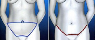
Swelling of the leg after vein surgery
After operations for varicose veins of the lower extremities, edema occurs in the early postoperative period. They are caused by damage to the soft tissues surrounding the venous vessel and lymphatic vessels. To prevent edema, phlebologists recommend that patients use elastic knitwear of the second compression class. Early activation and normal walking help normalize blood flow and prevent the development of edema.
Swelling after surgery on the veins more often occurs in patients who underwent surgery against the background of existing disturbances in the outflow of lymph from the lower limb or in the case of the development of erysipelas in the postoperative period. To reduce swelling of the lower limb, place it in an elevated position. Lymphatic edema decreases after applying a compression bandage with an elastic bandage. Pressure bandages are applied to certain areas of the legs. In the absence of the effect of conservative therapy, vascular surgeons perform minimally invasive interventions:
- Sewing of lymphatic vessels through the skin;
- Creation of channels in the subcutaneous tissue;
- Creating an outflow from the site of lymph accumulation.
To reduce swelling, phlebologists can perform suturing of lymphatic vessels throughout or in the wound.
Classification of hematomas
In modern medicine, when classifying hematomas, the following are taken into account:
- Relation to the vessel - pulsating and non-pulsating hematomas.
- Localization - in the cranial cavity, internal organs, under the skin or mucous membrane.
- The state of the blood in the affected area is suppurated, clotted, fresh, infected.
- Symptoms – limited, encysted, diffuse.
There are hematomas that do not fall into this classification. For example, intracerebral, intracranial, intraventricular. They are of the epidural or subdural type and cause serious complications.
1.General information
In medicine, a hematoma is a bruise (formed as a result of traumatic damage to small vessels), subcutaneous volumetric hemorrhage, or, to put it simply, an ordinary bruise. However, a lot depends on the exact area in which such a cluster formed and what its further dynamics are.
The tendency to hemorrhage into adjacent tissues is one of the distinctive features of trauma to the urinary system. Specific features of nephrotrauma also include severe pain syndrome (which is almost always observed with a kidney injury), certain difficulties with urination, and deterioration in the general condition of the victim. The body seems to be ringing the alarm bell: organs that should remain inviolable under any circumstances are damaged.
Kidney injuries, however, are the third most common among all documented injuries. Mechanical damage to this natural and irreplaceable blood filter is fatal in a significant percentage of cases. And a kidney hematoma, therefore, is not at all the same as a “regular” bruise.
A must read! Help with treatment and hospitalization!
Soft tissue hematomas
Soft tissue hematomas are divided into 3 types:
- Lungs - appear 24 hours after injury and are accompanied by mild pain. No special treatment is required.
- Medium - appear within 5-6 hours and are accompanied by pain and swelling. The motor function of the limb deteriorates. Consultation with a traumatologist is required.
- Heavy - formed within 2 hours after tissue damage. The function of the limb is impaired, acute pain and diffuse swelling are observed. You should immediately consult a doctor to determine a treatment strategy.
Immediately after the injury, swelling appears, and the skin acquires a purplish-bluish tint. After 5 days, the skin takes on a green tint as hemoglobin breaks down. Gradually, the hematoma resolves and “flows” down.
If there are no complications, the hematoma will resolve on its own. In the worst case, a hard area appears that causes discomfort and impairs motor function. When an intramuscular lump forms, external symptoms are rarely observed, but the limb swells significantly and an area forms inside, the touch of which causes severe pain.
Note! For chronic intramuscular hematomas, an MRI is prescribed to determine the location and extent of tissue damage.
When large lumps form, surgical intervention is required. Treatment is carried out by a traumatologist. The opening of infected seals is performed by a surgeon after a comprehensive diagnosis. The operation is performed on an outpatient basis, but for large hematomas hospitalization is required. An autopsy is performed, during which blood clots are removed and washing is carried out. Drainage and suturing are required. Sutures are not applied only for infected hematomas. Antibiotics are often prescribed in combination to eliminate the infection.
3. Symptoms, diagnosis
The most characteristic symptom of a kidney injury is intense specific pain, often radiating to adjacent areas (groin organs, intestines, chest) and caused by stretching of the renal capsule, acute local deficiency of blood supply (ischemia), mechanical pressure from the accumulated volume of blood, blocking the lumen of the ureter by coagulated blood clots . The purplish-bluish coloration of the skin in the lumbar region, edema, and swelling observed during external examination are not always found, and the absence of such visible signs does not exclude the presence of internal perinephric hemorrhage. A more characteristic symptom, which is also one of the main criteria for the severity of injury, is hematuria, i.e. the presence of blood in the urine (especially in the form of worm-shaped clots).
In clinical and diagnostic terms, the dynamics of the general condition is very important: in the first 1-2 days, symptoms can either gradually reduce or worsen into a life-threatening status with signs of sepsis and/or severe renal failure, unbearable pain, progressive hematuria, and shock.
In addition to collecting anamnestic information and complaints, examination and palpation (there are a number of diagnostically significant reactions), if a kidney hematoma is suspected, various methods of “examination from the inside” are used: cystoscopy, pyelography, angiography and other endoscopic, radiographic, ultrasound and tomographic studies, the choice of which is determined specific clinical situation.
About our clinic Chistye Prudy metro station Medintercom page!
Intracranial hematomas
Intracranial hematomas are divided into the following types:
- Epidural.
- Subdural.
- Intracerebral.
- Intraventricular.
Epidurals appear in 1-3% of cases and are due to injury to the middle meningeal artery. Pathology is often observed with skull fractures or depressed fractures. A hematoma develops in 2-3 hours or within 24 hours. Lack of treatment leads to coma. The first symptoms are confusion and weakness. Children rarely lose consciousness after a severe blow. Significant swelling of the brain does not lead to the detection of a light gap (which is rare in adults).
Subdurals appear in 1-7% of cases and pose a threat to human life, since death occurs in 60% of cases. There is an acute, subacute and chronic form of the pathology. Bleeding occurs due to a rupture of a vein or artery in the damaged area. People report nausea and severe headaches. Symptoms characteristic of compression of the brain stem are often observed. Lack of treatment and worsening symptoms lead to coma.
Intracerebral are observed extremely rarely with severe traumatic brain injuries. The light gap is not visible, the development of pathology occurs quickly. Hemiplegia or hemiparesis often occurs, as well as extrapyramidal symptoms.
Intraventricular diseases are rarely diagnosed due to the serious condition of patients. There are acute disturbances of consciousness, an increase in body temperature, a decrease in heart rate, and an increase in blood pressure. To establish a diagnosis, a survey of close people is carried out, since the patient is unconscious. To establish the location of the hematoma, MRI is used. In the most severe cases, lombal puncture is used.
Introduction
Despite the proven technique, operations for inguinal hernias can be accompanied by a number of intra- and postoperative complications (early and late), among which the most common are wound infection and hematomas (up to 10%), severe chronic postoperative pain (up to 3%) and relapse hernias (from 5 to 10%) [17]. Intraoperative complications also include direct damage to the structures of the groin area: intersection of a. epigastrica superficialis, a.
and
v.
femoralis, vv. Iliaca extern, Corona mortis; Vasa testicularis; strangulation followed by thrombosis
v.
femoralis ;
damage to Ductus deferens, N. ilioinguinlais, R. genitalis, N. genitofemoralis, N. iliohypogastricus
, intestine or bladder (the number of trocar injuries is 0.1-0.3%) [13, 18, 19, 22]. The incidence of early postoperative complications reaches 21.9% [3, 7, 8, 10, 12], among them long-term non-healing, including infected wounds, hematomas, seromas, swelling of the testicle and scrotum, acute pain, orchitis and orchiepididymitis [1, 2, 4, 6, 7, 18, 22].
The purpose of the study is to determine the dependence of the incidence of postoperative hematomas on the technique of performing inguinal hernia repair.
Material and methods
An analysis of the results of 625 planned and emergency hernia repairs performed for inguinal hernias and their complications in City Clinical Hospital No. 67 was carried out.
Moscow for the period from 2005 to 2012. In 150 (24%) cases, open plastic surgery of the posterior wall of the inguinal canal was performed, 115 of them made up the 1st group: in 23 (20%) - Bassini hernia repair was performed, in 67 (58.3%) - according to Shouldice, in 25 (21.7%) - according to Postempski. Patients who underwent emergency surgery when the strangulation lasted less than 6 hours were combined with patients who underwent elective hernia repair, since there were no significant differences in the technique and duration of the operations. 475 (76%) of 625 patients were operated on using a polypropylene prosthesis. Group 2 included 378 (60.5%) of 625 patients who underwent Lichtenstein type surgery. Patients in this group were divided into subgroups depending on the options for treating the hernial sac (HS): 127 (33.6%) operations without treatment of the hernia sac, 66 (17.5%) operations with reduction of the hernia sac and 185 (48.9%) operations with excision of the GM, and also depending on the type of reconstruction of the posterior joint: 179 (47.3%) patients received only prosthetics, 133 (35.2%) - prosthetics in combination with single-layer plastic surgery, 66 (17.5%) - with elements of multilayer plastic. Group 3 consisted of 83 (13.3%) of 625 patients who underwent laparoscopic transabdominal preperitoneal plastic surgery (Trans-Abdominal Preperitoneal - TAPP; n
=60) and total extra-abdominal plastic surgery (Total Extra-Peritoneal - TEP;
n
=23).
All groups of patients were comparable in terms of gender, age, presence of concomitant diseases, sides and timing of hernia, the presence of recurrent and inguinal-scrotal hernias, as well as previous operations on the lower abdomen. The results obtained were assessed retrospectively using primary documentation and prospectively: by recruiting our own subgroup without GM treatment, by telephone interviewing, and during control examinations. Calculations were carried out in the IBM SPSS Statistics 20.0.0 software environment using &khgr;2 tests, Fisher's exact test, Mann-Whitney U test, Kruskal-Wallis test, t
-test for comparison of means, binomial test, one-way analysis of variance;
Variables were checked for normality of distribution using the one-sample Kolmogorov-Smirnov test (values at p
≤0.05 were considered statistically significant).
results
Hemorrhagic complications in all groups were observed only in those patients who underwent operations with excision or reduction of the brain.
In group 1, scrotal hematocele was present in 4 (3.5%) patients (&khgr;2=2.361, p
=0.307), hematoma of the spermatic cord - in 5 (4.3%; &khgr;2=4.833,
p
=0.089), hemorrhagic infiltrate of the postoperative wound - in 2 (1.7%; &khgr;2=1.382,
p
=0.501) . Moreover, only 1 (4.5%) patient out of 22 who underwent emergency surgery developed a hematoma of the spermatic cord and a hematocele of the scrotum with a developed infiltrate, which was accompanied by partial divergence of the wound edges and prolonged lymphorrhea.
In group 2 (in the subgroup without reconstruction of the posterior joint), scrotal hematocele was noted in 9 (5.0%) cases (&khgr;2=2.118, p
=0.347), spermatic cord - in 6 (3.4%; &khgr;2=1.252,
p
=0.535), hemorrhagic infiltrate of the postoperative wound - in 5 (2.8%; &khgr;2=0.270,
p
=0.874) s subsequent lymphorrhea in 3 (1.7%) patients.
In the subgroup with single-layer plastic surgery of the PV (group 2), scrotal hematocele and spermatic cord hematoma were noted in 1 (0.8%) patient (&khgr;2=1.303, p
=0.521), and the ultrasound picture indicated imbibition of the testicle with blood;
hemorrhagic infiltrate of the postoperative wound - in 4 (3.0%; &khgr;2=5.333, p
=0.070), lymphorrhea - in 5 (3.8%; &khgr;2=2.082,
p
=0.353).
In the subgroup with multilayer plastic surgery of the glaucoma (group 2), operations were performed mainly without treatment of the brain (in 59 of 66 patients). No hemorrhagic complications were observed in this subgroup.
The results of endoscopic operations were compared with the results of open prosthetics. In the 3rd group, 4 (4.8%) patients developed hematomas of the cervical cavity left in the inguinal canal (when compared with observations of a hematocele of the scrotum after open prosthetics, Fisher’s exact test with a two-sided p
=0.283).
In 2 of these 4 cases, a transinguinal approach with excision of the cervical cavity and debridement of the hematoma was required. In 4 (4.8%) patients, an inguinal hematoma was observed that required evacuation (Fisher's exact test with a two-sided p
= 0.109).
In 2 (2.4%) patients, infiltration was noted in the area of trocar punctures with subsequent delayed wound healing (Fisher's exact test with a two-sided p
= 1.000).
There were no significant differences in the frequency of hemorrhagic complications in patients of groups 1, 2 and 3, which corresponds to literature data, however, there was a natural tendency to reduce the number of complications when minimizing surgical aggression in relation to the cerebral hemorrhage and spermatic cord, as well as when performing functional reconstruction of the vertebral joint. .
Discussion
Hematoma is one of the most common complications of inguinal hernia repair, its incidence is 1-16.8% [15, 21, 22]. From a pathogenetic and practical point of view, we distinguish 6 types of hematomas as complications of inguinal hernia repair.
First type
— hematoma of a postoperative wound, or rather imbibition of the wound edges with blood. The reasons for this complication are insufficient hemostasis during surgery and rough traction, and both the first and second can be a consequence of technical difficulties during surgery (depth of the wound, obesity of the patient, the presence of adhesions in recurrent and/or irreducible hernia, fragility of blood vessels in atherosclerosis, drug-induced hypotension and the absence of obvious bleeding during surgery, taking antiplatelet agents and anticoagulants, hemophilia), insufficient qualifications, as well as the choice of surgical technique [11, 13, 14, 16, 18-20, 22]. The latter is becoming increasingly important with the development of new accesses. Thus, with a standard transinguinal approach with an incision length of up to 8 cm, which creates sufficient exposure, the chance of making technical errors, as well as the occurrence of hematomas, is apparently less than when trying to reduce the length of the surgical wound.
The development of single access surgery also faces problems associated with excessive port pressure on the edges of a single surgical wound of the anterior abdominal wall. Thus, the frequency of trocar hernias with the SI (single incision) technique is 2-3% [4], which is comparable to the recurrence rate after open hernia repair with prosthetics for inguinal hernia.
Second type
, which can be separately isolated, is a hematoma of the spermatic cord or its imbibition with blood. Surgeries for indirect inguinal hernia should be noted as a risk of this complication, since in this case surgeons traditionally dissect the membranes of the spermatic cord when isolating the inguinal hernia [6]. Further processing of the GM, to one degree or another, aggravates the risk of bleeding, although the safest option is to reduce the GM into the abdominal cavity [9]. Partial excision of the GM, intersection at the neck, gathering with sutures, dissection and suturing in the form of an additional layer - all these technique options are fraught with additional trauma, one of the complications of which may be the development of bleeding and hematoma [3, 7].
Hematomas of the third type
have no consequences as they resolve - these are hematomas (ecchymoses) of the skin of the scrotum, which are sometimes difficult to distinguish from another variant of hematomas - scrotal hematomas
(type four)
.
Theoretically, the fourth type can be divided into two subtypes: hematocele of the scrotum (Fig. 1, a and further in the color insert)
and hematoma of the testicle itself
(Fig. 1, b)
, which are probably the result of the same complication - hematoma of the testicle cord.
Figure 1. Ultrasonograms.
a — hematocele of the scrotum. The arrows indicate the walls of the scrotum; b - imbibition of the testicle with blood (on the left - frontal, on the right - sagittal plane). The arrows indicate the contours of the testicle. Fifth type
- hematoma of the preperitoneal space, occurs, as a rule, during manipulations within the preperitoneal space, both with transinguinal preperitoneal plastic surgery and with endoscopic approaches, in which technical difficulties of hemostasis arise, as well as when isolating the GM and spermatic cord in oblique, irreducible and inguinal scrotal hernias.
Thus, during endoscopic interventions, bleeding may occur if the epigastric artery is damaged by a trocar inserted without proper visual control (Fig. 2)
.
Figure 2. Access stage for total extraperitoneal hernioplasty.
Multiple branches of the inferior epigastric artery (1) and vein (2) are visible - Corona mortis It should be noted that preperitoneal hematomas, as a rule, proceed more safely, in clinically erased forms, but giant preperitoneal hematomas also occur. M. Parvaize et al. [18] reported the development of a hematoma with a volume of 770 ml after open right-sided inguinal hernia repair in a 71-year-old patient taking dipyridamole.
In Fig. 3
another type of hematoma (
sixth
) is shown after inguinal herniotomy - hematoma of the cervical cavity, left after its intersection in the scrotum.
Figure 3. Hematoma of the hernial sac left in the inguinal canal after endoscopic surgery.
1 - hernial sac; 2 - spermatic cord. The stage of re-operation (from the transinguinal approach) is presented - isolation of the GM left in the scrotum during TARR for a large right-sided indirect inguinal hernia (type IV according to Nyhus). A repeat operation was performed due to the development of intestinal obstruction in the patient in the postoperative period, caused by the introduction of a loop of the small intestine into the “access window” in the peritoneum due to the failure of the sutures, while the hematoma of the severed GM, left in the scrotum, simulated a recurrent hernia (so called “false relapse" [5]. The GM was excised, then repeated laparoscopy was performed, intestinal obstruction was eliminated and the peritoneal defect was re-sutured (no signs of failure of the SG of the PC were noted after prosthetics). It is usually possible to differentiate seromas and hematomas during ultrasound examination based on the nature of the liquid component (it is more “dense” in hematomas and contains inclusions - blood clots), although clinically these complications are not always distinguished [15].The only reliable diagnostic method is the evacuation of the liquid contents.
The above classification of hematomas, as well as research data, in our opinion, should help in assessing the complications that arise when using one or another method of hernia repair.
Thus, the immediate and long-term results of emergency and planned inguinal hernia repair using autoplasty methods, abdominal wall prosthetics during open and endoscopic surgery do not depend on gender, age, concomitant diseases, but are associated with the technical features of performing a number of surgical techniques (excessive discharge, excision of the hernial sac , refusal to reconstruct the posterior wall of the inguinal canal during its prosthetics). The occurrence of traumatic complications of inguinal hernia repair, such as hematomas of the spermatic cord and scrotum, orchitis, orchiepididymitis, is directly dependent on the technique of treating the hernial sac, and the number of relapses depends on the option of reconstruction of the posterior wall of the inguinal canal, the reliability of which increases when prosthetics is supplemented with autoplasty techniques.
Identification of classification characteristics of the most common complications of inguinal hernia repair (in particular, hematomas) facilitates the assessment of the influence of various techniques and techniques on the results of operations.
Diagnostics
A hematoma is diagnosed by visual examination. If the hemorrhage is located deep under the skin, in internal organs, or in a joint, it is often very difficult to assess its size and possible consequences.
Patients are prescribed an examination, which may include:
- Ultrasound of internal organs, joints;
- computed tomography and magnetic resonance imaging;
- puncture (puncture with a needle): for example, a puncture of the knee joint is often done if there is a suspicion that blood has accumulated in it after an injury.
Thromboembolic complications of phlebectomy
Swelling of the leg after vein surgery can occur due to deep vein thrombosis. This complication of the postoperative period develops under the influence of the following risk factors:
- Increased blood clotting after surgery;
- Injuries of venous vessels;
- Temporary physical inactivity.
Since after surgery on the veins there is swelling of the lower limb and pain, doctors often do not recognize thrombosis of the deep arteries of the leg in time. Phlebologists at the Yusupov Hospital always adhere to the rule: if after phlebectomy swelling and other signs of impaired venous outflow increase, thrombosis of the deep veins of the leg should be excluded. For this purpose, patients undergo ultrasound scanning using color and power Doppler. To prevent thromboembolic complications, patients are prescribed acetylsalicylic acid, dosed compression and an active motor regimen.
Edema due to deep vein thrombosis of the lower extremities can develop even after the patient is discharged home. Sometimes patients do not attach importance to this, and they develop pulmonary embolism. To prevent this from happening, if your leg swells after vein surgery, immediately call the contact center of the Yusupov Hospital. Before the ambulance arrives, you should take a horizontal position, place a pillow under your leg, apply an elastic bandage or put on compression stockings and take an aspirin tablet.
Treatment
Minor bruises can be treated conservatively: physiotherapeutic procedures and medications are prescribed.
In case of large accumulations of blood, the hematoma is treated surgically: it is opened, the blood or pus is evacuated, washed with antiseptics and drainage is installed. Antibiotics are prescribed if necessary.
When hemorrhaging into internal organs, it is often necessary to perform surgical intervention, during which it is necessary not only to remove the spilled blood, but also to stop the bleeding.
The multidisciplinary CELT clinic employs experienced traumatologists and surgeons who perform operations on hematomas of various locations. Modern techniques used in our clinic help provide effective treatment and minimize the risk of complications.
General complications
As a result of surgical intervention, systemic disorders occur in the body, which are considered as postoperative complications:
- painful sensations. They are relieved with analgesics, antispasmodics and desensitizing agents in various combinations;
- nervous system disorders. If the patient suffers from insomnia, he is prescribed sleeping pills and sedatives;
- Postoperative bronchitis and pneumonia appear more often in smokers. In such cases, antibiotics and symptomatic therapy are prescribed;
- acute heart failure is considered the most dangerous complication requiring measures to save the patient;
- acute embolism and thrombosis in cardiovascular pathologies, increased blood clotting, varicose veins. To prevent such complications, it is necessary to place the operated limbs above the level of the body, tighten the feet and legs with elastic bandages, and prescribe therapy with anticoagulants and desagrenants;
- complications of the gastrointestinal tract in the form of stomatitis and sialadenitis (inflammation of the salivary glands) or more serious consequences of the operation - paresis (lack of tone and peristalsis) of the stomach and intestines;
- On the part of the bladder, difficulty and retention of urination are often observed. Catheterization may help;
- Bedsores form when the patient remains in one position for a long time in a supine position. To prevent them, good patient care is needed. When bedsores appear, they are treated with antiseptic solutions and wound healing agents.
Treatment of complications after surgery is a very important point in the rehabilitation program for a surgical patient. This is given due attention by surgeons at the Sanmedexpert clinic. As a result, the number of postoperative complications is minimized.
Orthopedics and traumatology services at CELT
The administration of CELT JSC regularly updates the price list posted on the clinic’s website. However, in order to avoid possible misunderstandings, we ask you to clarify the cost of services by phone: +7
| Service name | Price in rubles |
| Appointment with a surgical doctor (primary, for complex programs) | 3 000 |
| X-ray of the chest organs (survey) | 2 500 |
| Ultrasound of soft tissues, lymph nodes (one anatomical zone) | 2 300 |
All services
Make an appointment through the application or by calling +7 +7 We work every day:
- Monday—Friday: 8.00—20.00
- Saturday: 8.00–18.00
- Sunday is a day off
The nearest metro and MCC stations to the clinic:
- Highway of Enthusiasts or Perovo
- Partisan
- Enthusiast Highway
Driving directions